Intravenous pyelogram
Jump to navigation
Jump to search
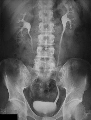
An intravenous pyelogram (IVP) is an x-rayexamination of the kidneys, uretersand urinary bladderthat uses iodinated contrastmaterial injected into veins.An x-ray (radiograph) is a noninvasive medical test that helps physicians diagnose and treat medical conditions. Imaging with x-rays involves exposing a part of the body to a small dose of ionizing radiationto produce pictures of the inside of the body. X-rays are the oldest and most frequently used form of medical imaging.When a contrast material is injected into a vein in the patient's arm, it travels through the blood stream and collects in the kidneyand urinary tract, turning these areas bright white. An IVP allows the radiologistto view and assess the anatomy and function of the kidneys, ureters and the bladder.An intravenous pyelogram examination helps the physician assess abnormalities in the urinary system, as well as how quickly and efficiently the patient's system is able to handle waste.The exam is used to help diagnose symptoms such as blood in the urine or pain in the side or lower back.The IVP exam can enable the radiologist to detect problems within the urinary tract resulting from:
kidney stones
 enlarged prostate
tumors in the kidney, ureters or urinary bladder
kidney stones
 enlarged prostate
tumors in the kidney, ureters or urinary bladder