Imaging Talar Fractures
Introduction
Talus fractures can present a challenge to the radiographer both radiographically and in terms of image interpretation. They also present a challenge to the referring doctor who often isn't sure whether to ask for ankle radiography or foot radiography (the talus seems to belong to both!)... of course, asking for imaging of the talus would appear to be eminently sensible.
This page considers all aspects of plain film imaging of the talus.
Anatomy
<a class="external" href="http://www.jaaos.org/cgi/content/full/13/8/492/JA0004404FIG1" rel="nofollow" target="_blank"> </a>
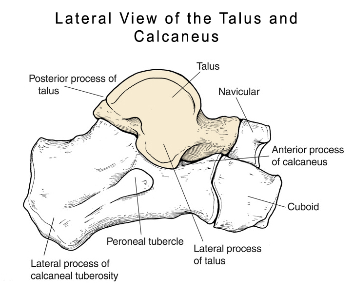
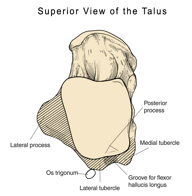
<a class="external" href="http://www.joint-pain-expert.net/talus-fracture.html" rel="nofollow" target="_blank">source: Talus Fracture: Diagnosis and Treatment (18/1/2011)</a>Talus fracture can involve different parts of the bone which may be
- neck
- body
- head
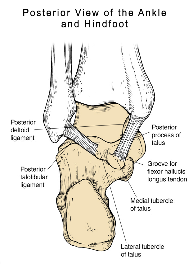
- process
adapted from <a class="external" href="http://www.jaaos.org/cgi/content/full/13/8/492/JA0004404FIG1" rel="nofollow" target="_blank">Process and Tubercle Fractures of the Hindfoot
Berkowitz and Kim J Am Acad Orthop Surg.2005; 13: 492-502 (18/1/2011)</a>Talus - Receives forces 5 – 10 times the body weight during normal ambulation
- 3/5 of surface is covered by articular cartilage
- Articular Cartilage is 1 – 2 mm thick
<a class="external" href="http://www.wheelessonline.com/ortho/Talus_Osteochondral_Lesions" rel="nofollow" target="_blank">http://www.wheelessonline.com/ortho/Talus_Osteochondral_Lesions (18/1/2011)</a>
adapted from <a class="external" href="http://www.jaaos.org/cgi/content/full/13/8/492/JA0004404FIG1" rel="nofollow" target="_blank">Process and Tubercle Fractures of the Hindfoot
Berkowitz and Kim J Am Acad Orthop Surg.2005; 13: 492-502 (18/1/2011)</a>
adapted from <a class="external" href="http://www.jaaos.org/cgi/content/full/13/8/492/JA0004404FIG1" rel="nofollow" target="_blank">Process and Tubercle Fractures of the Hindfoot
Berkowitz and Kim J Am Acad Orthop Surg.2005; 13: 492-502 (18/1/2011)</a>
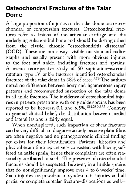
Osteochondral Fractures of the Talar Dome
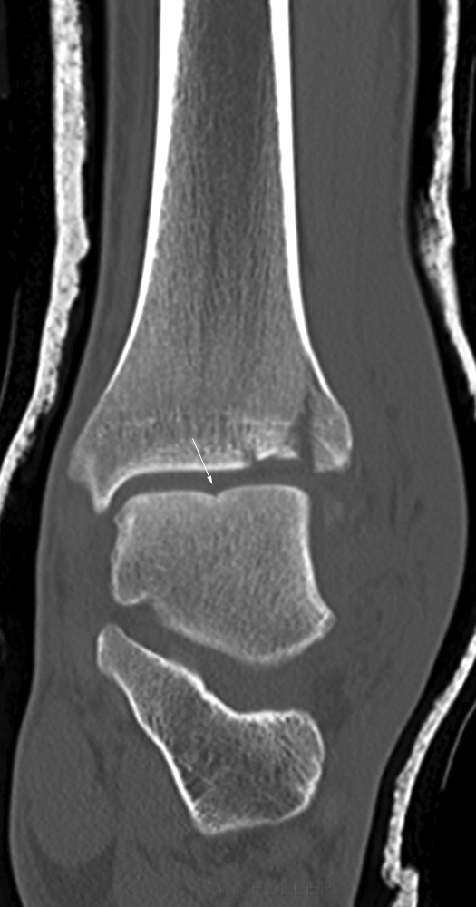
This quote from Bruce Browner et al provides an overview of osteochondral fractures of the talar dome. Talar dome fractures can be demonstrated on plain films of the ankle but can easily be missed and/or simply not demonstrated. Follow up radiographs can reveal these fractures because of the secondary changes in the subchondral bone. MRI and CT can be useful in demonstrating these fractures in an acute setting.
Hawkin's Sign
The Hawkin’s Sign:
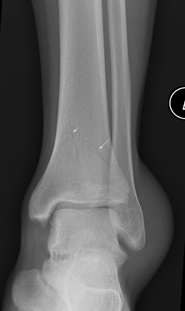
- A subchondral radiolucent band in the talar dome at 6-8 weeks post injury, seen on an AP-mortice view of the ankle
- Represents re-vascularisation of talar body, and sufficient blood supply to allow for disuse osteopaenia from resorption
- Absence is worrisome for AVN
<a class="external" href="http://thebonesurgeon.com/Documents/Talus+Fractures.pdf" rel="nofollow" target="_blank">http://thebonesurgeon.com/Documents/Talus%20Fractures.pdf</a> 18/1/2011
Fractures of the Posterior Process of the Talus
Case 1
Case 2
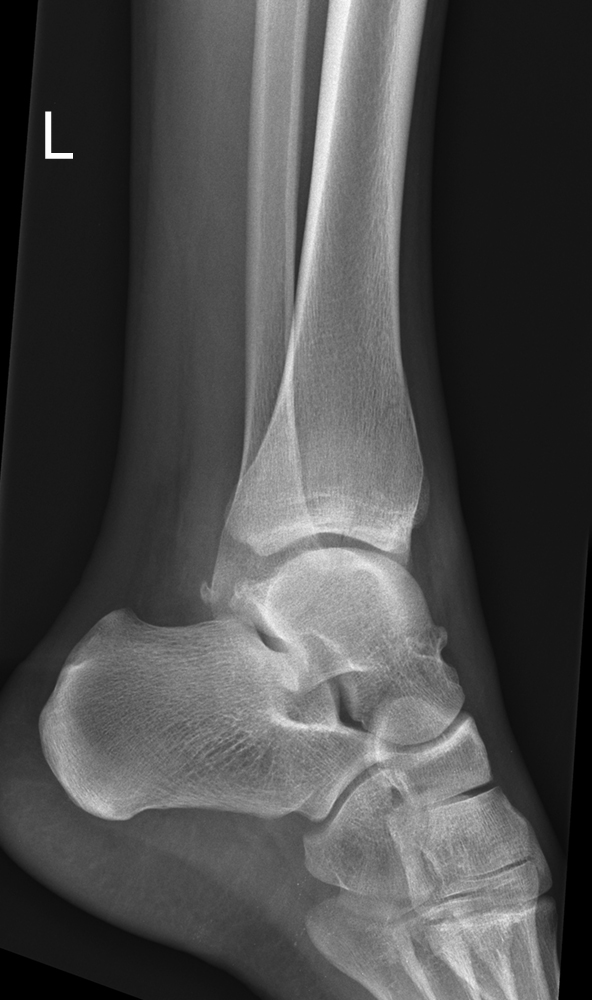
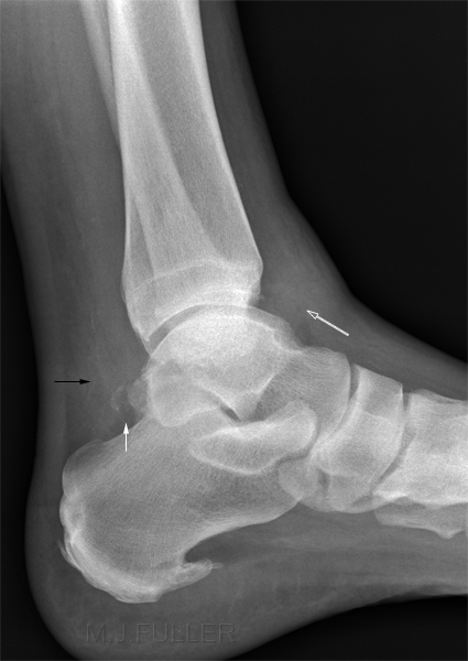
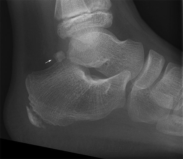
This 10 year old boy presented to the Emergency Department with a history of several months of pain in the area of the Achilles tendon insertion into the calcaneum. He was referred for foot radiography which the radiographer interpreted as calcaneum radiography.
The arrowed structure is either an accessory ossicle known as Os trigonum, or a fracture of the posterior process of the talus. Whilst there is some abnormal adjacent fluid density within Kager's fat pad, the clinical signs favour this structure being an Os trigonum. Irrespective of the correct diagnosis, the difficulty in distinguishing between Os trigonum and a posterior talus fracture is evident.
"Fractures of the os trigonum- The os trigonum is an accessory bone (sesamoid) located posterior to the posterior tubercle of the talus. It is present in 5-14% of the population and is frequently unilateral. This accessory bone may be fused to the talus, calcaneus, or both. Patients with a fracture of the os trigonum often give a history of having sustained an ankle sprain weeks to months earlier, at which time radiographs were interpreted as normal. They give a history of persistent posterior and posterolateral ankle pain, swelling, and giving way of the ankle. All had a 25° decrease in plantar flexion of the ankle and pain to palpation posterior to the tibia but anterior to the Achilles tendon. The pain was enhanced by forced plantar flexion of the ankle or resisted plantar flexion of the great toe."
Qouted from
<a class="external" href="http://emcrit.org/030-064/051-ankle.foot.htm" rel="nofollow" target="_blank">Emergency Medicine and ED Critical Care
http://emcrit.org/030-064/051-ankle.foot.htm</a>
... back to the Wikiradiography home page
... back to the Applied Radiography page
