Imaging Rib Fractures
The identification of rib fractures radiographically is a controversial subject. Rib radiography is considered unnecessary in many centres because it often has, at best, a marginal effect on patient management. It should also be considered that undisplaced rib fractures can be missed on plain film images. Irrespective of which viewpoint you subscribe to, if you are planning to undertake rib radiography you should be attempting to achieve the highest image quality at the lowest radiation dose. This page considers rib radiography techniques.
Mechanism of Injury
- Trauma
- Coughing
- Pathological
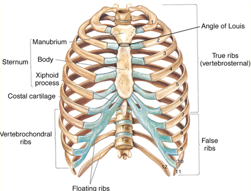
Anatomy
The Appropriateness of Rib Radiography
The ACR suggest that rib radiography is more appropriate in children than adults. They note the following.
| <a name="1292">"While rib fractures can produce significant morbidity, the diagnosis of associated complications (such as pneumothorax, hemothorax, pulmonary contusion, atelectasis, flail chest, cardiovascular injury, and injuries to solid and hollow abdominal organs) may have a more significant clinical impact. Radiographs are specific but not very sensitive (for undisplaced fractures), and clinical examination is sensitive but not specific."</a> | ||
| [[While rib fractures can produce significant morbidity, the diagnosis of associated complications (such as pneumothorax, hemothorax, pulmonary contusion, atelectasis, flail chest, cardiovascular injury, and injuries to solid and hollow abdominal organs) may have a more significant clinical impact. Radiographs are specific but not very sensitive (for undisplaced fractures), and clinical examination is sensitive but not specific.|National Guidelines Clearing House]] | ||
Counterpoint- arguments in favour of Rib Radiography
Point Comment Fractures of the first rib are associated with mediastinal/vascular injury. This contention is not universally supported by research Fractures of the lower ribs are associated with trauma to the abdominal viscera These patients should be considered for CT imaging A flail chest may not be clinically evident in some patients, particularly obese patients patients with ribs fractures identified radiographically are more likely to receive appropriate pain relief identification of multiple rib fractures can have management implications rib fractures in children have high association with NAI
Technique Notes
- A PA chest image should be included with rib views to assess for pneumothorax (erect, expiration)
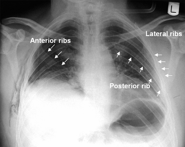
- It is difficult to include all ribs in a single exposure- centre to include the symptomatic anatomy
- specific below diaphragm exposure technique may be required
- including the lung apices may demonstrate pneumothorax
- roll the scapula away from the lungfield where possible
- upper ribs are best demonstrated on deep inspiration and lower ribs on deep expiration
- low kVp's should be employed; 60 - 65 kVp for upper ribs and 70 - 75 kVp for lower ribs (digital radiography kVps can be higher)
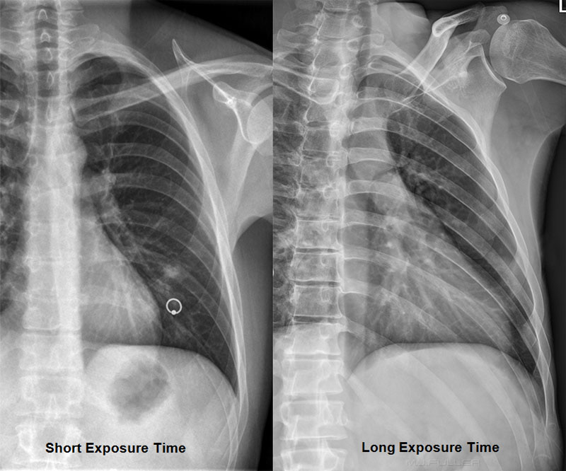
- A long exposure time in a compliant patient can produce very high quality results.
- Long exposure time rib views should not be attempted in the erect position (movement unsharpness)
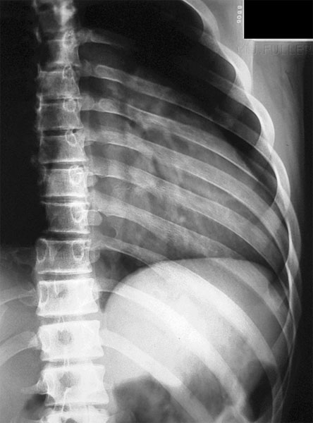
Long Exposure Time technique
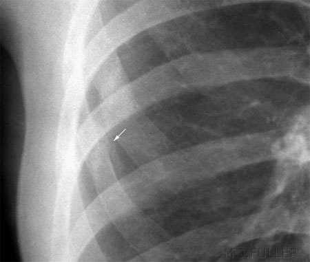
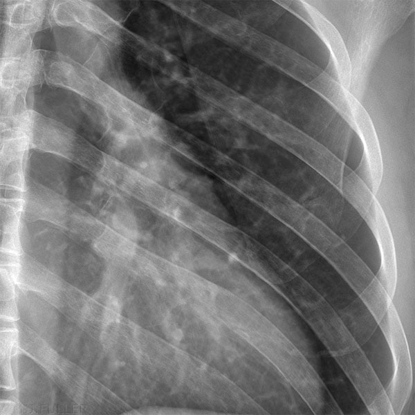
Old vs New Rib Fractures
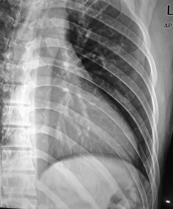
This image demonstrates an old rib fracture. The presence of callus new bone formation around the fracture indicates that it is a healing fracture.
Fractured Ribs on Other Views
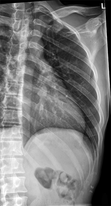
Film-Screen Technique
DR Imaging
Sonography
Ultrasound diagnosis of rib fractures can be very effective. One study which compared rib radiography with ultrasound imaging of patients with acute rib injuries reported the following;
"At presentation, radiographs revealed eight rib fractures in six ( 12%) of 50 patients and sonography revealed 83 rib fractures in 39 (78%) of 50 patients. Seventy-four (89%) of the 83 sonographically detected fractures were located in the rib, four (5%) were located at the costochondral junction, and five (6%) in the costal cartilage. Repeated sonography after 3 weeks showed evidence of healing in all re-examined fractures. Combining sonography at presentation and after 3 weeks, 88% of subjects had sustained a fracture."
<a class="external" href="http://www.ajronline.org/cgi/reprint/173/6/1603" rel="nofollow" target="_blank">J. F. Griffith et al
Sonography Compared with Radiography in Revealing Acute Rib Fracture.
AJR:173, December 1999</a>
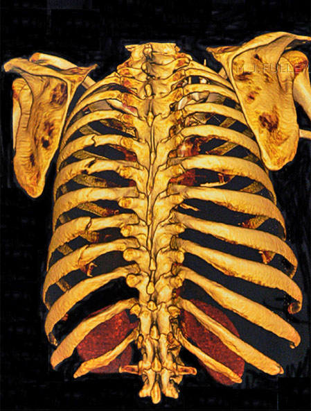
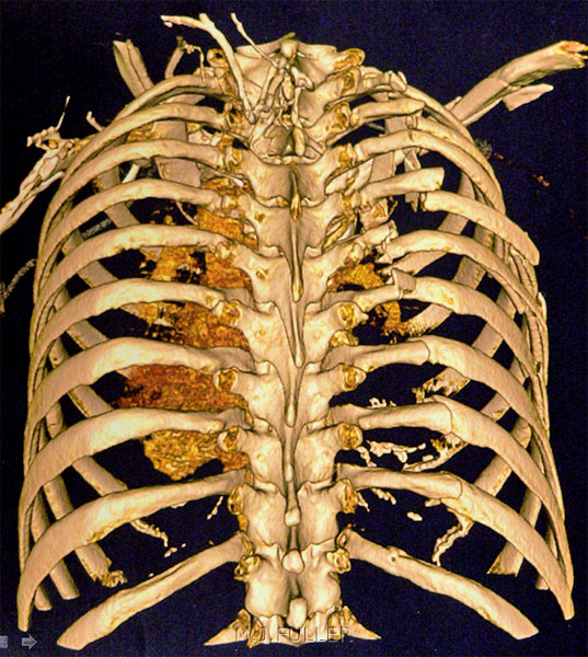
CT Imaging
...back to the Wikiradiography home page
...back to the Applied Radiography home page
