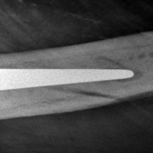
Imaging Periprosthetic Hip Fractures
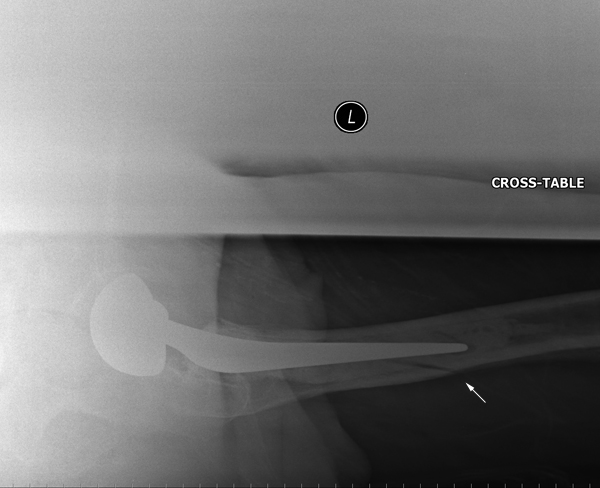
RadiographyWith increases in the number of hip replacement operations in many countries, the number of periprosthetic femur fractures can be expected to increase. Minimally displaced periprosthetic fractures can be difficult to detect. Radiographers should be aware of the possibility of periprosthetic fractures and the radiographic techniques required to demonstrate them.
<a class="external" href="http://biomed.papers.upol.cz/pdfs/bio/2004/01/13.pdf" rel="nofollow" target="_blank">Leopold Pleva, Milan Šír, Roman Madeja
OUR EXPERIENCES WITH THE TREATMENT OF PERIPROSTHETIC FRACTURES OF FEMUR
Biomed. Papers 148(1), 75–79 (2004)</a>Elderly patients who have fallen onto their hips are commonly referred for hip radiography with a provisional diagnosis of neck of femur fracture. It is the author's experience that these patients are frequently not examined carefully by the referring doctor and can have a hip prosthesis on the side of interest. The radiographer should examine the patient priot to hip radiography in elderly patients. This will take very little time and may change the radiographer's approach to the imaging.
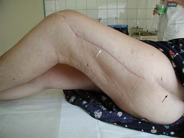
This patient has evidence of previous hip surgery (surgical scar- black arrow) and surgical treatment of a periprosthetic fracture (surgical scar- white arrow).
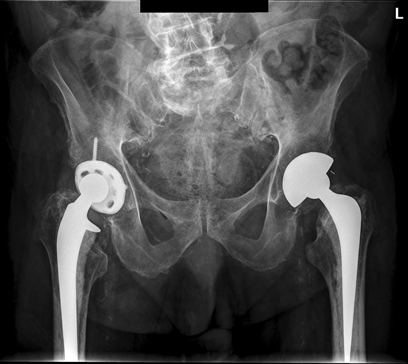
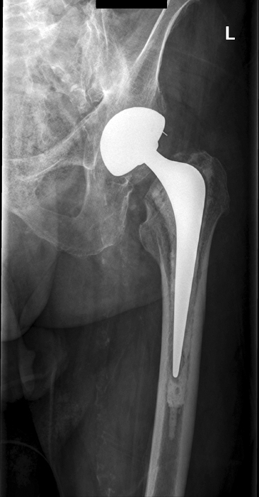
When the history of hip surgery is established, it is important to radiographically asses the entire femur.
Case 1
