Imaging Mandibular Fractures
Introduction
Mandible fractures are commonly seen in Emergency Departments. Imaging mandible fractures often raises the question of whether the imaging should include plain films, OPG or both. This page considers all aspects of plain film imaging of mandible fractures.
OPG or Plain Films?
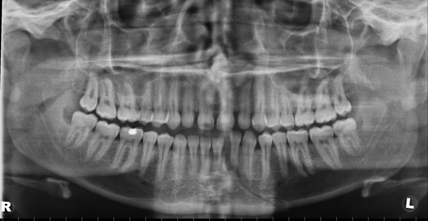
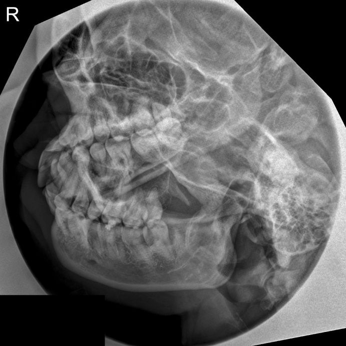
Access to an OPG machine is undoubtedly useful in departments that treat patients with mandible trauma. The question arises as to which is the superior imaging approach- plain film or OPG? You could argue that an OPG is a type of tomogram and tomography should never be the first line of imaging. Equally there are cases where the OPG will demonstrate fractures clearly that are not seen on plain films. Conversely, the opposite is also true.
Should the OPG be the first line of investigation (like a screening tool) and the plain films employed as supplementary imaging or should the plain films be taken first and OPG employed to clarify any equivocal findings? The success of the plain films is also operator dependant- with a reduction in requests for plain film imaging of the skull and facial bones, many junior radiographers are not confident about their skull/facial bone plain film imaging skills.
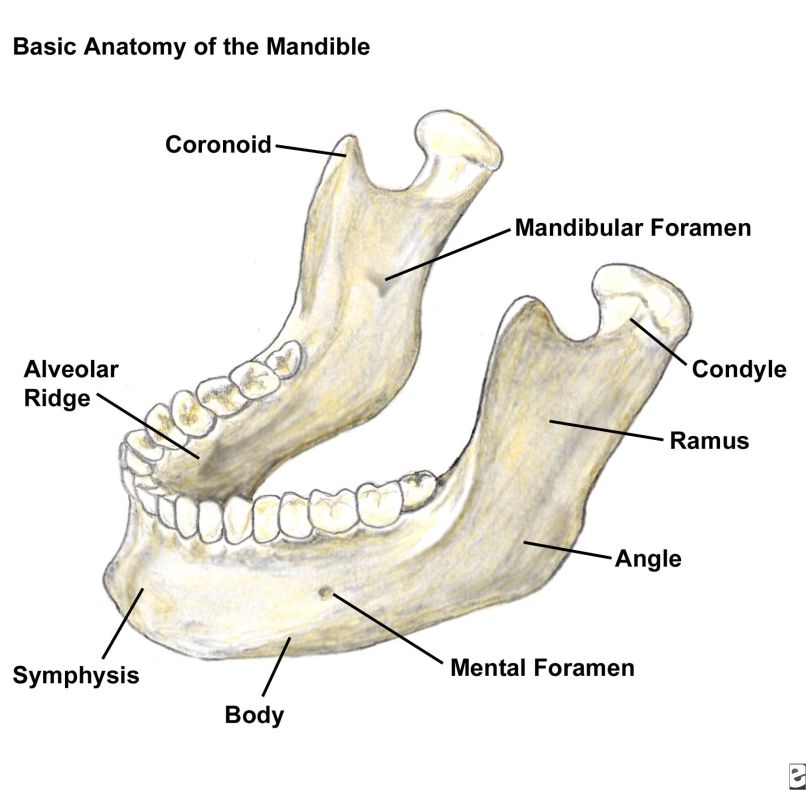
Anatomy
<a class="external" href="http://emedicine.medscape.com/article/391549-overview" rel="nofollow" target="_blank">http://emedicine.medscape.com/article/391549-overview</a>
Case Study 1
Comment
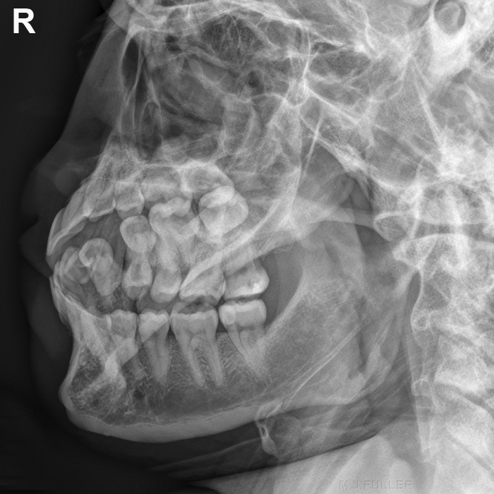
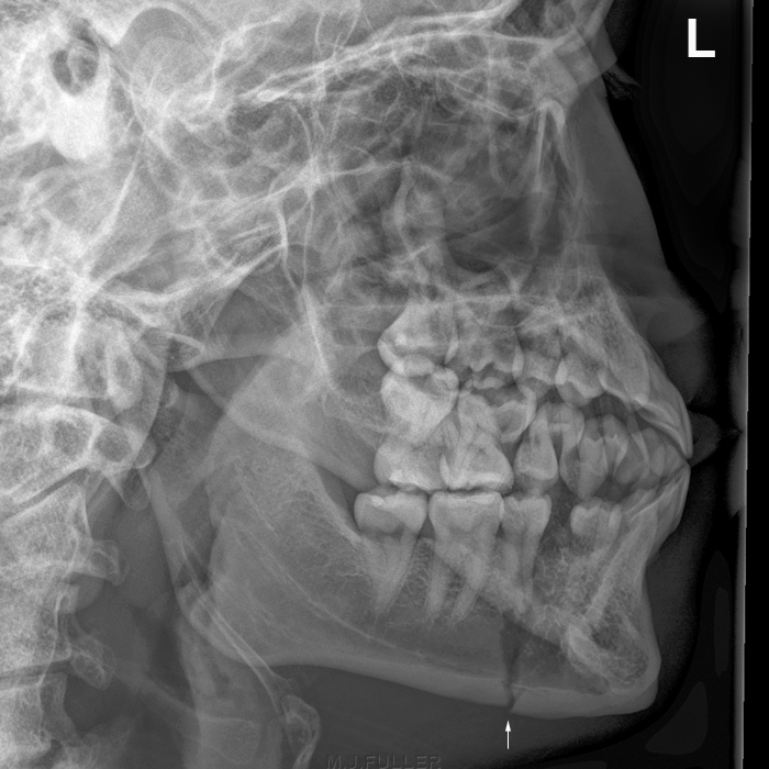
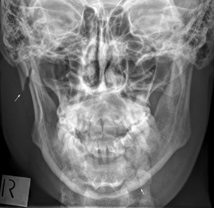
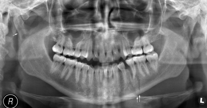
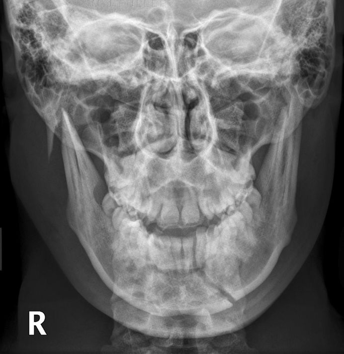
This case demonstrates the limitations of the OPG in demonstrating mandible fractures. Although both fractures are visible, the right fracture is not obvious and the nature of the right ramus fracture (separation and displacement) is not clearly demonstrated. By contrast, the right ramus fracture could not be missed on the PA view image and the orientation, separation of fragments and displacement are clearly shown.
Case Study 2
.... back to the wikiradiography home page
... back to the Applied Radiography home page
