Introduction Abdominal hernias are a common abdominal pathology. They range between asymptomatic and surgical emergencies. This page is dedicated to the plain film imaging of abdominal hernias.
Definition An abdominal hernia is an abnormal protrusion or extension of intra-abdominal contents through an internal or external foramen or space
Types of Hernias Abdominal hernias are divided into internal and external. Internal hernias are uncommon in developed countries and are very difficult to diagnose on abdominal plain film. Internal hernias that contain small bowel may present as a small bowel obstruction. External hernias are a common cause of small bowel obstruction
| Internal | Comment |
- Left and right paraduodenal fossae
|
|
- Hernia through defect in lower omentum
|
|
- Hernia into lesser sac through foramen of Winslow or through a defect in the gastrocolic ligament
| - small bowel gas may be visible adjacent to the stomach
- a supine decubitus lateral of the upper abdomen may be useful
|
| |
| |
| - This could be considered a type of diaphragmatic hernia
|
| External |
|
| | - can easily be missed if the obturator foramen are not included in the image
|
| | - a supine decubitus tangential of the hernia lump can reveal incarcerated bowel. An Aluminium filter technique is very usefully employed.
|
| | - occurs at site of previous surgery
- can be very large and inoperable if longstanding
- a supine decubitus tangential of the hernia lump can reveal incarcerated bowel. An Aluminium filter technique is very usefully employed.
|
- Spigelian(or lateral ventral hernia)
| - hernia through the spigelian fascia
|
Radiography Successful radiography of abdominal hernias starts with good patient communication. It is surprising how often you uncover a key piece of clinical information (that the referring doctor has failed to include in the referral) when talking to the patient. A simple question like "how are you?" can lead to all manner of relevant information e.g. "I'm really worried about this lump on my tummy". This is a highly relevant piece of information when all the referring doctor has offered is "abdo pain, ? SBO". From a patient care point of view, this is also beneficial- the patient will be more relaxed in the knowledge that you are diligent in your approach and understand of his/her problem.
The radiography of abdominal hernias does not differ greatly from other acute abdominal imaging except for a few important considerations.
Inguinal and femoral Hernias.
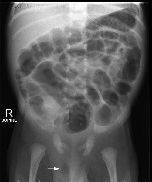 | It is "unnatural" for Radiographers to deliberately and knowingly irradiate the scrotum, particularly a baby's scrotum. Despite this radiographer discomfort, if a baby is suspected to have an inguinal or femoral hernia, it will be missed if the scrotum is not included on the abdominal image.
If your departmental policy is to asses all potential inguinal/femoral hernias in babies by ultrasound imaging, the problem is avoided.
This baby has an inguinal/femoral hernia that is evident from the pocket of gas in the scrotum(white arrow). If the radiographer had centred a little higher, the hernia could have been missed |
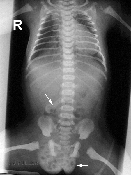 | This baby has an inguinal hernia with air-filled bowel within the scrotum (bottom arrow)
The artifact is an umbilical clamp (arrow) |
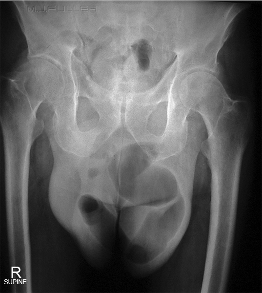 | It is unlikely that a scrotal hernia of this size would be missed by the referring doctor on clinical examination. The patient is usually referred for "abdominal" imaging to assess whether the hernia is causing a bowel obstruction and to assess whether the hernia contains bowel. Clearly, to assess whether the hernia contains bowel the scrotum will need to be included in the image. This can be a little awkward for the patient and radiographer if the patient is uncomfortable about telling you about the size of their scrotum (understandable). A careful, considered and professional approach will usually pay dividends.
This scrotum clearly contains air-filled small bowel (no arrows required!). |
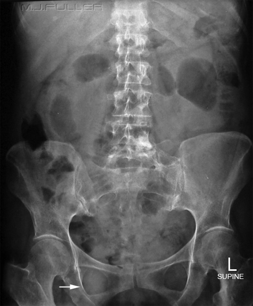 | Occasionally the only evidence of inguinal/femoral hernia on abdominal plain film is an uneven density of the obturator foramen. It has been argued that all acute abdominal plain film imaging should include the obturator foramen for this reason. My understanding is that this is not taught in schools of radiography, and perhaps rightly so. The benefits of detecting these hernias would have to be weighed against the increased gonad dose to the vast majority of patients who don't have a hernia.
At a more realistic and practical level, where the referring doctor/radiographer suspects inguinal/femoral hernia, inclusion of the obturator foramen would be justified. |
|
|
Para-umbilical, incisional, ventral hernias
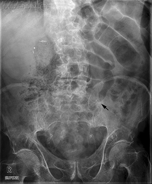 | This patient presented with acute abdominal pain. The patient had a history of previous umbilical hernia and an incisional hernia was evident on physical examination.
The radiographer performed an AP supine abdominal examination which showed evidence of small bowel obstruction. A possible point of entry of the small bowel into the incisional hernia was also identified (black arrow).
The radiographer considered that the diagnosis of SBO caused by incarcerated small bowel in the incisional hernia was likely, but additional confidence in the diagnosis would be achieved if a tangential view demonstrated small bowel gas in the hernia (see below) |
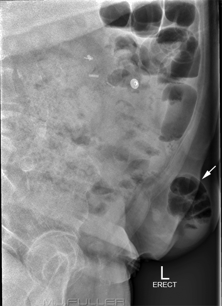 | The radiographer identified the position of the patient's hernia and performed an erect tangential view of the hernia. The hernia is clearly demonstrated, as are the small bowel loops within the hernia (white arrow).
The tangential image was performed without an aluminium filter. The images were acquired using a Philips Digital Diagnost DR machine (release I). After acquisition, the images were post-processed to achieve maximum latitude (Gamma adjustment). This quality of image is unlikely to be achieved with film/screen or CR imaging. The use of an aluminium filter will improve success rates if not using DR.
The tangential view is arguably best performed in the supine decubitus position. The reasoning is that incarcerated bowel could be missed if it contains no air. The air is more likely to rise into the incarcerated bowel if it is the least dependent part of the bowel i.e. with the patient supine. |
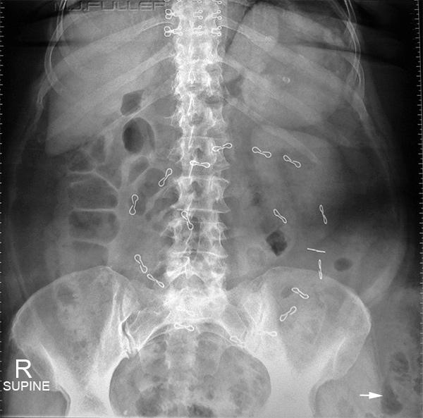 | This patient has multiple staples from previous abdominal surgery. There is bowel gas seen lateral to the left hemi-pelvis. This bowel gas in likely to be contained within an incisional hernia. This appearance can also occur in morbidly obese patients with pendulous abdomens.
Close coning of the primary radiation field in patients with possible external hernias is contra-indicated. |
Incisional Hernia
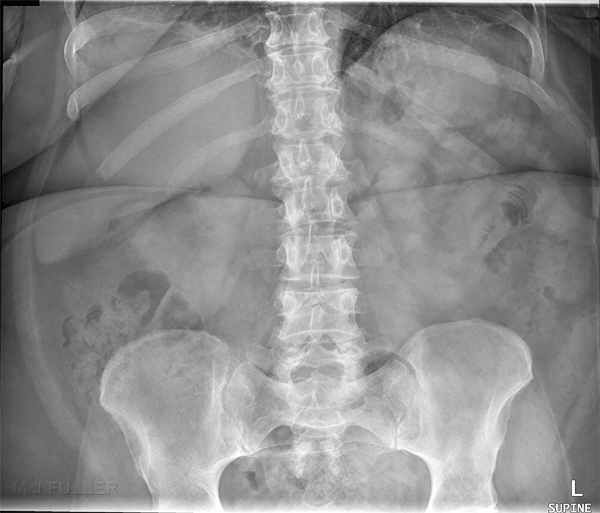 | This patient presented to the Emergency Department with acute abdominal pain. She had a history of an incisional hernia which had been surgically repaired twice.
The radiographer asked the patient about her symptoms and she identified a para-umbilical palpable lump just above and to the left of her umbilicus. The radiographer noticed that the lump became visible when she moved onto the X-ray table. This was presumed to be caused by increased abdominal pressure associated with moving onto the X-ray table from the barouche.
The supine abdominal image demonstrated a general paucity of intestinal gas, particularly in the large bowel- this can indicate the presence of intestinal obstruction. There is a suggestion of several fluid filled loops of small bowel with a solitary suspect looking air-filled loop on the left. This loop was considered suspect given that it is
- air-filled,
- it is minimally dilated
- it demonstrates a coiled spring stretched appearance
|
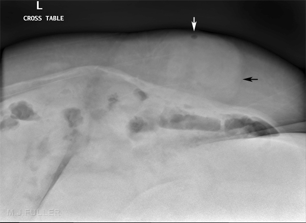 | Given the possibility of incarcerated small bowel within the presumed incisional hernia, it was considered prudent to perform a tangential view of the hernia. The patient was positioned in the supine position and a supine decubitus projection was employed to try and demonstrate the hernia. To improve the chances of success, the patient was positioned slightly RPO and the patient was asked to increase abdominal pressure (it was previously noted that the hernia became more prominent with a slight increase in abdominal pressure). This also helped to ensure that the hernia was imaged in profile.
An aluminium filter was employed in an attempt to deal with the considerable variation in tissue thickness to be imaged.
The images were acquired using a Philips Digital Diagnost DR machine.
The incisional hernia is demonstrated as a rounded soft tissue density within the subcutaneous tissue (black arrow). Note that this density is positioned between the muscle of the patient's anterior abdominal wall and the skin.
Importantly, there is a small air bubble (white arrow) within the hernia which is likely to be a small air collection within small bowel (this could be considered a single pearl of the string-of-pearls sign).
When you put together the patient's history, the patient's clinical signs, the paucity of large bowel gas, the suspect loop of small bowel on the left, the soft tissue density demonstrated on the supine decubitus view, and the gas within this density, you have a likely diagnosis of
"recurrence of incisional hernia with associated small bowel obstruction".
The patient proceeded to theatre for surgical reduction of the hernia.
Importantly, the patient did not have a CT scan of the abdomen prior to surgery despite the fact that the service was readily available at the time.
Even more importantly, the radiographers involved in the plain film examination believed that their diligence provided the surgeons with a confident plain film diagnosis without the need for CT imaging. |
Summary Successful demonstration of external abdominal hernias will require radiographers to undertake dedicated supplementary views in some patients. Talking to the patient about their presentation may reveal a history of hernia not mentioned by the referring doctor. A clear and comprehensive knowledge of the patient's clinical signs and history may precipitate important supplementary radiographic views. Successful demonstration of incarcerated bowel in an external hernia could conceivably save the patient from undergoing an abdominal CT and expedite treatment of their condition.
This type of clinical approach to radiography is more effective and more satisfying that a purely photographic approach.
...back to the Applied Radiography home page


