Hepatic-Portal Venous Gas
Introduction
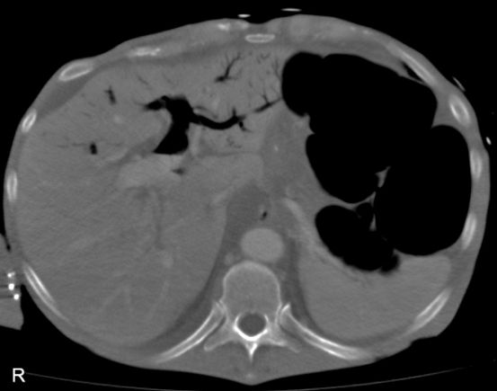
Plain film demonstration of Hepatic-Portal Venous Gas (HPVG) can be seen in patients of all ages including premature babies. Although the clinical significance is variable and somewhat ill-defined, it is common for this appearance to represent a surgical emergency. This page considers all aspects of the plain film demonstration of HPVG. Not unlike toxic megacolon, HPVG is not a disease but rather a sign of potentially serious acute abdominal pathology.
Synonyms
portal vein gas, pneumoportogram, gas embolisation of the portal vein
Causes
Causes of HPVG Cause Cases Necrotic bowel 72 Ulcerative colitis 8 Intra-abdominal abscess 6 Small Bowel Obstruction 3 Gastric Ulcer 3
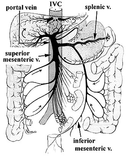
Anatomy of the Portal System
<a class="external" href="http://application.fnu.ac.fj/classshare/Medical_Science_Resources/MBBS/MBBS1-3/PBL/Liver/Anatomy+of+the+Liver-Univ+of+Queensland/portalv.JPG" rel="nofollow" target="_blank">
</a><a class="external" href="http://application.fnu.ac.fj/classshare/Medical_Science_Resources/MBBS/MBBS1-3/PBL/Liver/Anatomy+of+the+Liver-Univ+of+Queensland/portalv.JPG" rel="nofollow" target="_blank">source: Gross Anatomy of the Liver
University of Queensland</a>The portal system includes all the veins which drain the blood from the abdominal part of the digestive tube (with the exception of the lower part of the rectum) and from the spleen, pancreas, and gall-bladder. From these viscera the blood is conveyed to the liver via the portal vein. In the liver this vein ramifies like an artery and ends in capillary-like vessels termed sinusoids, from which the blood is conveyed to the inferior vena cava by the hepatic veins. <a class="external" href="http://www.bartleby.com/107/174.html" rel="nofollow" target="_blank">http://www.bartleby.com/107/174.html</a>
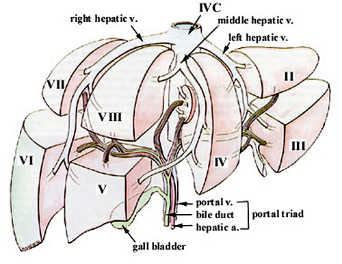
Blood flow to the liver is unique in that it receives both oxygenated and deoxygenated blood. As a result, the partial pressure of oxygen (pO2) and perfusion pressure of portal blood are lower than in other organs of the body. Blood passes from branches of the portal vein through cavities between hepatocellular sinusoids. Blood also flows from branches of the hepatic artery and mixes in the sinusoids to supply the hepatocytes with oxygen. This mixture percolates through the sinusoids and collects in a central vein which drains into the hepatic vein. The hepatic vein subsequently drains into the inferior vena cava.
<a class="external" href="http://radiopaedia.org/articles/portal-venous-system" rel="nofollow" target="_blank">quoted from http://radiopaedia.org/articles/portal-venous-system</a>
<a class="external" href="http://application.fnu.ac.fj/classshare/Medical_Science_Resources/MBBS/MBBS1-3/PBL/Liver/Anatomy+of+the+Liver-Univ+of+Queensland/liverseg.JPG" rel="nofollow" target="_blank">Source: http://application.fnu.ac.fj/classshare/Medical_Science_Resources/MBBS/MBBS1-3/PBL/Liver/Anatomy%20of%20the%20Liver-Univ%20of%20Queensland/liverseg.JPG</a>"The liver is divided into 8 segments, each with its own vascular inflow, outflow and bile drainage. The segments may be pictured as wedge-shaped units, with a broad base forming the diaphragmatic surface and an apex pointing towards the IVC posteriorly. The liver segments are defined by the hepatic veins which form the boundaries, while the portal vein hepatic artery and bile ducts run through the centre of the segments. Segment(s) can therefore be removed by resection along segment boundaries."
<a class="external" href="http://application.fnu.ac.fj/classshare/Medical_Science_Resources/MBBS/MBBS1-3/PBL/Liver/Anatomy+of+the+Liver-Univ+of+Queensland/" rel="nofollow" target="_blank">quoted from </a><a class="external" href="http://application.fnu.ac.fj/classshare/Medical_Science_Resources/MBBS/MBBS1-3/PBL/Liver/Anatomy+of+the+Liver-Univ+of+Queensland/" rel="nofollow" target="_blank">
Gross Anatomy of the Liver
University of Queensland</a>
Pathophysiology
Liebman et al (1978) reviewed 60 published cases with 4 cases of their own and reported the following
[P.R. Hepatic--portal venous gas in adults: etiology, pathophysiology and clinical significance. Annals of Surgery , v.187(3); Mar 1978]
Plain Film Appearance
Hepatic-portal Venous Gas
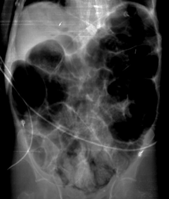
<img align="bottom" alt="portal venous gas" src="http://image.wikifoundry.com/image/1/jGWwjd2Ap2NdY6Fd58WtVA240521" title="portal venous gas"/>This is a magnified view of the erect abdominal plain film RUQ. The hepatic portal venous gas is clearly seen as well as intramural gas (black arrow) and surgical sutures (white arrow).
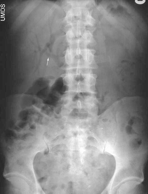
The radiographic appearance of HPVG has been described as a branching radiolucency which can almost extend to the liver capsule.
The appearance is sufficiently characteristic in appearance that it should not be confused with the appearance of gas within the biliary tree.Gas in the Biliary Tree The radiographic appearance of gas in the biliary tree is usually sufficiently distinctive to avoid confusion with HPVG. This patient has gas in the biliary tree with gas-contrast demonstration of the bile duct, pancreatic duct, and left and right hepatic branches.
Case 1
... back to Wikiradiography home page
back to the Applied Radiography home page
