Heart
Jump to navigation
Jump to search
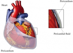
Is a thin double walled sac that surrounds the heart and contains the roots of the great vessels it is characterised by two layers.
Starting in the right atrium, the blood flows through the tricuspid valve to the right ventricle. Here it is pumped out the pulmonary semilunar valve and travels through the pulmonary artery to the lungs. From there, blood flows back through the pulmonary vein to the left atrium. It then travels through the mitral valve to the left ventricle, from where it is pumped through the aortic semilunar valve to the aorta. The aorta forks, and the blood is divided between major arteries which supply the upper and lower body. The blood travels in the arteries to the smaller arterioles, then finally to tiny capillaries which feed each cell. The (relatively) deoxygenated blood then travels to the venules, which coalesce into veins, then to the inferior and superior venae cavae and finally back to the right atrium where the process began.
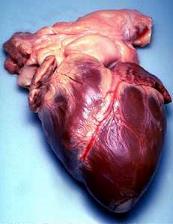
| The heart is a muscular organ in all vertebrates responsible for pumping blood through the blood vessels by repeated, rhythmic contractions. The heart is composed of cardiac muscle, an involuntary muscle tissue which is found only within this organ. The average human heart beating at 72 Beats per minute (BPM), will beat approximately 2.5 billion times during a lifetime spanning 66 years. It is usually situated in the middle of the thorax with the largest part of the heart slightly offset to the left (although sometimes it is on the right, see dextrocardia). The heart is usually felt to be on the left side because the left of the heart (left ventricle) is stronger (it pumps blood to all body parts). The left lung is smaller than the right lung because the heart occupies more of the left hemithorax. The heart is enclosed by a sac known as the pericardium and is surrounded by the lungs. The pericardium comprises two parts: the fibrous pericardium, made of dense fibrous connective tissue; and a double membrane structure containing a serous fluid to reduce friction during heart contractions (the serous pericardium). The mediastinum, a subdivision of the thoracic cavity, is the name of the heart cavity | Heart removed from human during autopsy <a class="external" href="http://upload.wikimedia.org/wikipedia/commons/b/b7/Humhrt2.jpg" rel="nofollow" target="_blank">  </a> </a> |
Pericardium
Is a thin double walled sac that surrounds the heart and contains the roots of the great vessels it is characterised by two layers.
- The first layer is an outer fibrous pericardium which anchors the heart to the surrounding structures.
- The second layer is the inner serous pericardium, consisting of an outer parietal layer and inner visceral layer.
Fibrous Pericardium
- Flask shaped bag with the neck of which is closed by its fusion with external coats of the greater vessels.
- The base of the fibrous pericardium is is attached to the central tendon and to the muscular fibers of the left side of the diaphragm.
- The diaphragmatic attachment consists of loose fibrous tissue, which can be easily broken down, unlike the fused portion of the sac. Above, the fibrous pericardium not only blends with the external coates of the great vessels, but is continuous with the pretrachael layer of the deep cervical fascia.
- It is also attached to the posterior surface of the sternum by the superior and inferior stenopericardiac ligaments; the upper portion passing to the manbrium and the lower portion passing to the xiphoid process.
Serous Pericardium
- Parietal layer
- Lines the inner surface of the fibrous pericadium
- Visceral layer (Serous Pericardium and also known as Epicardium)
- Covers the outside of the heart
- These two layers are continuous with each other and reflect along and attach to the great vessels.
- The thin space separating them is called the pericardial cavity, which contains serous fluid that to lubricate the membranes and facilitate the almost frictionless continuous movement of the heart.
Heart wall
The heart is consists of three layers that are very distinctive: an external epicardium, a middle myocardium, and an internal endocardium. Arrangement of cardiac muscle in the heart wall permits the compression necessary to pump large volumes of blood out of the ventricles.
Heart Muscles - Resemble skeletal fibers because they are striated, with extensive capillary networks.
- Contract as a single unit; muscle impulses are distributed simultaneously throughout all fibers either of the atria or the ventricles
- Specialized cell-cell contacts;"intercalated discs"
- Arranged in spiral bundles and wrapped around and between the heart chambers
- Differences
- Sacroplasmic reticulum is less extensive
- No terminal cisternae
- Lacks tight associations of smooth endoplasmic reticulum and transverse tubules
- Differences
Chambers
The Heart externally is composed of four hollow chambers. Two smaller atria and two larger ventricles.- Atria (Atrium)
- Thin-walled chambers located superiorly
- The anterior part of each atrium is a wrinkled, flap-like extension called the auricle
- Separated from ventricles externally by deep coronary sulcus that extends around the heart.
- All sulci house blood vessels that supply the heart
- All sulci house blood vessels that supply the heart
- Receives blood returning to the heart from both the circulatory circuits
- Right atrium receives blood from the systemic circuit
- Left atrium receives blood from the pulmonary circuit .
- Blood passing through the right atrium enters the right ventricle, which are the inferior chambers
-
Pulmonary Trunk is one of the two large arteries. It carries the right ventricle into the pulmonary circuit -
Aorta the second large artery. Conducts blood from the left ventricle into the systemic circuit
Heart valves
Right atriventricular valve
Between right atrium and right ventricle
Prevents backflow of blood into right atrium when ventricles contract
Pulmonary semilunar valve
Between right ventricle and pulmonary trunk
Prevents back flow of blood into right ventricle upon ventricle relaxationLeft atriventricular valve
Between left atrium and left ventricle
Prevents back flow into left atrium upon ventricle contractionAortic semilunar valve
Between left ventricle and ascending aorta
Prevents back flow of blood into left ventricle upon ventricle relaxation
Functioning
The function of the right side of the heart is to collect de-oxygenated blood, in the right atrium, from the body and pump it, via the right ventricle, into the lungs (pulmonary circulation) so that carbon dioxide can be dropped off and oxygen picked up (gas exchange). This happens through the passive process of diffusion. The left side of the heart collects oxygenated blood from the lungs into the left atrium. From the left atrium the blood moves to the left ventricle which pumps it out to the body. On both sides, the lower ventricles are thicker and stronger than the upper atria. The muscle wall surrounding the left ventricle is thicker than the wall surrounding the right ventricle due to the higher force needed to pump the blood through the systemic circulation.
Starting in the right atrium, the blood flows through the tricuspid valve to the right ventricle. Here it is pumped out the pulmonary semilunar valve and travels through the pulmonary artery to the lungs. From there, blood flows back through the pulmonary vein to the left atrium. It then travels through the mitral valve to the left ventricle, from where it is pumped through the aortic semilunar valve to the aorta. The aorta forks, and the blood is divided between major arteries which supply the upper and lower body. The blood travels in the arteries to the smaller arterioles, then finally to tiny capillaries which feed each cell. The (relatively) deoxygenated blood then travels to the venules, which coalesce into veins, then to the inferior and superior venae cavae and finally back to the right atrium where the process began.
Fibrous Skeleton
The fibrous skeleton is located between the atria and ventricles, formed by dense irregular connective tissue. Functions- Separating atria and ventricles
- Anchoring the heart valves
- Provide electrical insulation between atria and ventricles (prevents all of the heart chambers from beating at the same time)
- Provides framework for attachment of cardiac muscle
