Hand/Wrist
Jump to navigation
Jump to search
This page will introduce student radiographers to the movements of the wrist and fingers. As well as the prominent land marks of the distal upper extremities.
Go Back to Surface Anatomy Home Page
Hand & Wrist Surface Anatomy | Other pages of interest |
This page will introduce student radiographers to the movements of the wrist and fingers. As well as the prominent land marks of the distal upper extremities.
Movements Of the Wrist:
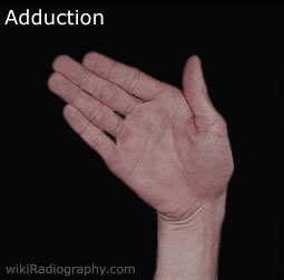
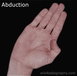
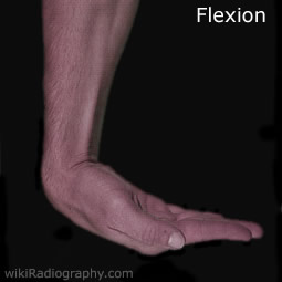
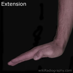
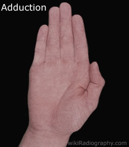
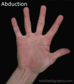
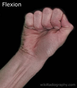
| | The wrist is essentially a double row of small short bones, called carpals, intertwined to form a malleable hinge. This joint allows four movements of the wrist to be performed - Adduction, Abduction, Flexion and Extension shown in the images to the left. When using a combination of these movements the wrist is said to undergo circumduction (circular movement). Adduction is when the wrist is deviated towards the ulna. Abduction is when the wrist is deviated towards the radius. Flexion is when the joint angle at the wrist is decreased. Extension is when the joint angle at the wrist is increased. | |
| |
Movements of the Fingers:
| | Each hand has five fingers or digits. Normally humans have five digits on each hand. Polydactyly is a condition where there is more than five digits and hypodactyly is a condition where there is less. Aside from the genitals, the fingertips possess the highest concentration of touch receptors and thermoreceptors among all areas of the human skin, making them extremely sensitive to heat, cold, pressure, vibration, texture, and moisture. Thus fingers are commonly used as sensory probes to ascertain properties of objects encountered in the world, and so they are prone to injury. When using digits they are numbered 1 through 5, starting at the thumb.
Often on radiology request forms, referring doctors use the digit system of numbering, this however can lead to confusion and it is best practice to ask the patient which is the affected finger. (It is very easy for the referring physician to accidentally start counting from the wrong side the hand!) The fingers are able to undergo four types of movement - Adduction, Abduction, Flexion and Extension. When using a combination of these movements they undergo circumduction or circular movement. Adduction - a movement of the fingers closer to the sagittal plane. Abduction - a movement of the fingers away from the sagittal plane. Flexion - when the joint angles of the fingers decrease. Extension - when the joint angles of the fingers increase. |
| | |
| |
Anatomical Snuff Box:
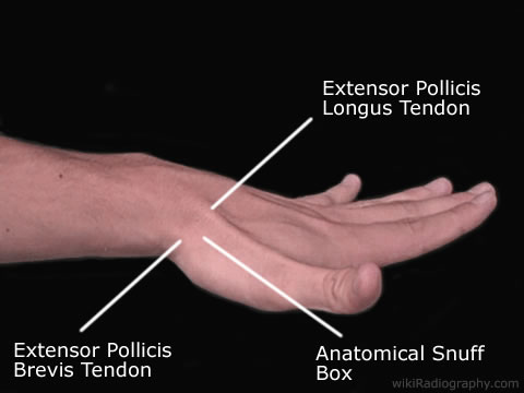
| | The anatomical snuff box is a region the radiographer needs to become acquainted with. The floor of the snuffbox is primarily the scaphoid bone. This is a common fracture site when falling on an outstretched hand (FOOSH). If tender when palpated, it may indicate that there is a fracture of the scaphoid bone. Gentle palpation or having the patient point to the sorest point in the wrist may help you decide if dedicated scaphoid images need to be performed. The lateral border of the snuff box is made by the extensor pollicis brevis tendon, the medial border by the extensor pollicis longus tendon. It is most visible and more pronounced during thumb extension. |
Palmer Surface:
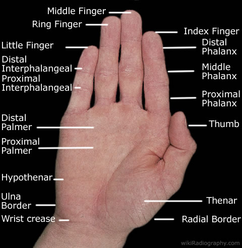
| | The anterior surface of the hand is often refered to as the palmar surface. The crease of the wrist is where the joint undergoes flexion and extension. The intrinsic muscle groups are the thenar and hypothenar muscles (thenar referring to the thumb, hypothenar to the small finger). All the other muscles that provide flexion and extension of the fingers are located in the forearm. The fat that is located on the fingers and palm of the hand is more malleable and does not rebound after being compressed compared to fat located elsewhere on the body. Presumably because it is better suited to moulding around surfaces when picking up objects The thumb contrasts with the other four fingers by being the only finger that
Each of the phalanges are palpable. There is three in each finger and as mentioned two in the thumb. |
Dorsal Surface:
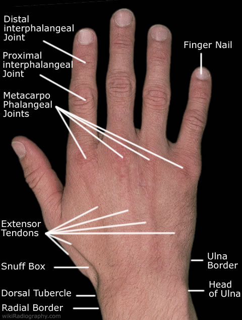
| | The posterior surface of the hand is often referred to as the dorsal surface. On the radial side of the wrist just below the snuff box the dorsal tubercle can be felt. The tubercle serves as a trochlea or pulley for the extensor pollicis longus tendon. On the ulna side of the wrist the ulna head can be palpated. It is extremely difficult to feel the ulna styloid. On the dorsal surface you can palpate all five metatarsals of the hand. You should be able to 1) Feel each of the metatarsal bases above the carpal bones. 2) Feel the long shaft of each metatarsal. 3) Feel each metatarsals biconvex head (around the MCP joint) The metacarpophalangeal joints (MCP) can be felt and are often referred to in layman's terms as the knuckles. Each of the phalanges are palpable. There is three in each finger and two in the thumb. At the end of each finger is a nail. Each fingernails is made of a tough protein called keratin and are produced from living skin cells in the fingers. There is no nerve endings in the fingernails and they serve to protect the ends of the fingers. |
Go Back to Surface Anatomy Home Page
