Terminology- FRD (Focal Spot to Receptor Distance)
- ORD (Object to Receptor Distance)
- OFD (Object to Focal Spot Distance) or object-film distance
- SID (Source Image-receptor distance)
- IR (Image receptor)
3 Dimensions Displayed in 2 Dimensions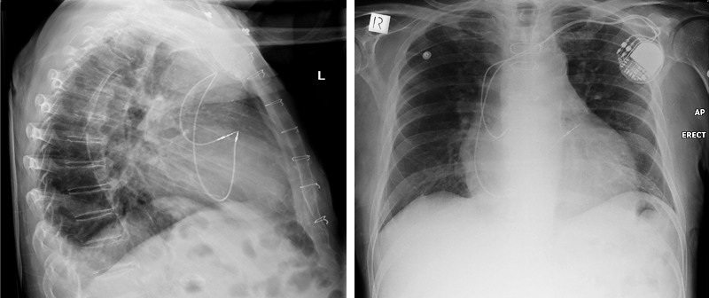 | An X-ray image is a representation of a 3 dimensional object (the patient) in two dimensions (the image). This provides a common and recurring dilemma in plain film image interpretation.
The lateral chest (left) suggests that the tips of the patient's pacemaker leads are touching. This may or may not be the case. The only correct statement you can make on the basis of the lateral image alone is that the pacemaker lead tips are superimposed in this projection. The AP erect chest image demonstrates that the tips of the pacemaker leads are separated considerably. |
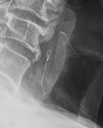 | This patient presented to the Emergency Department with a possible foreign body in the upper oesophagus. The lateral projection demonstrated an apparent metallic wire foreign body in the upper oesophagus. The position of the wire cannot be confirmed on this single projection (the wire could be external to the patient). A second projection at 90 degrees to the lateral would confirm the position of the apparent foreign body.
Note also the appearance of a knot in the wire. |
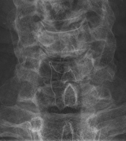 | The AP projection image demonstrated the wire near the midline suggesting that this was in fact a metallic foreign body lodged in the upper oesophagus.
Noe that the appearance of a knot in the wire was not real. |
MagnificationAll anatomy demonstrated on a plain film image has some degree of magnification. The degree of magnification will depend on geometric factors. Whilst this might appear to be something that you learn to pass your radiography exams, it can have surprisingly important implications in the real world. Some examples are shown below.
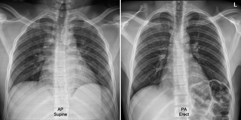 | The left image was taken in the resus room with the patient on a dedicated X-ray resus trolley/gurney/barouche. The FFD/SID/FRD was limited by the tube height (?radiographer height/reach) and the OID is greater than normally present with a table bucky setup. These geometric factors result in an abnormally wide cardiac/mediastinal shadow on plain film supine chest imaging. The right image is a PA erect chest image on the same patient. This technique demonstrates that the mediastinum is not abnormally wide.
This becomes a problem in patients who have presented to the ED following severe trauma such as MVA. They are usually imaged in the supine position- supine chest radiography is the only technique option. Whilst the apparently widened mediastinum is likely to be geometric (radiologists like to call this projectional), the mediastinum appears widened on the best (only) available imaging. This cannot be ignored by the clinicians and the patient is often referred for CT chest imaging. |
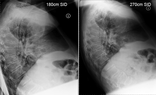 | Both images (left) are taken with the patient sitting in an identical position on a barouche/trolley. The patient could not be moved closer to the IR to reduce radiation dose and geometric magnification. At 180 cm SID (source-image receptor distance) the radiographer experienced difficulty including all of the chest anatomy. The right image is taken with the patient in exactly the same position with the SID increased to 270cm- this is not a routinely employed SID but can be usefully employed where geometric magnification is likely to cause difficulties.
Pleural effusion (left hemithorax), osteopenia and old wedge fractures noted. |
... back to the Applied Radiography home page ... back to the Wikiradiography home page 


