Foot - DP
Jump to navigation
Jump to search
Radiographic Positioning
| Adult Foot - Dorsiplantar Projection | Other related pages of interest |
| Name of Projection | Foot - Dorsiplantar (DP) |
| Area Covered | Phalanges, metatarsals, navicular, cuneiforms and cuboid |
| Pathology Shown | Fractures, dislocation, foreign body, joint space abnormalities |
| Radiographic Anatomy | Foot Radiographic Anatomy |
| IR Size & Orientation | 24 x 30 cm Portrait, divided in two can usually fit 2 views, use lead masking for unused area |
| Film / Screen Combination | Detail (CR and DR as recommended by manufacturer) |
| Bucky / Grid | No |
| Filter | Yes - when using film a thin filter covering phalanges and distal metatarsals |
| Exposure | 55 kVp 3.2 mAs |
| FFD | 100cm |
| Central Ray | Centre to include foot Directed at base of 3rd metatarsal
|
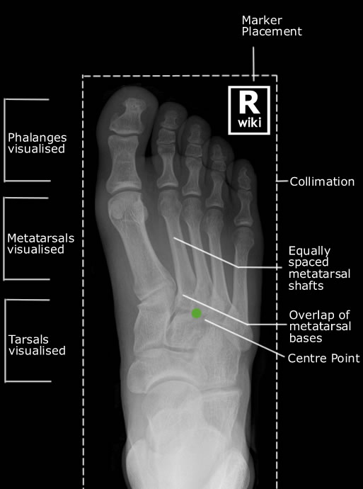
| Collimation | Outer skin margins of foot on four sides |
| Markers | Distal and Lateral Marker orientation AP |
| Shielding | Gonadal (check your department's policy guidelines) |
| Respiration | Not applicable |
| Positioning |
|
| Critique | Positioning
|
| Special Notes | Effect of lateral rotation
Effect of medial rotation
|
Foot DP - Evaluation
