Finger Radiography
Introduction
<a class="external" href="http://pagnal.wordpress.com/2009/12/03/climbing-grades/" rel="nofollow" target="_blank">
http://pagnal.wordpress.com/2009/12/03/climbing-grades/</a>I once overheard a colleague say "a finger injury isn't important until it's yours". The same could be said of finger radiography. There are some common mistakes in finger radiography technique that can be easily avoided.
The hand is a human being's most exquisite organ of direct interaction with the surrounding universe
Skeletal trauma: basic science, management, and reconstruction Bruce D. Browner, Alan M. Levine, Jesse B. Jupiter, Peter G. Trafton, Christian Krettek
Anatomy
Overview
- PIP joint consists of medial and lateral condyles on the proximal phalanx, with matching concavities on the associated distal phalanx.
- IP joints have flexion and extension movements , but they are relatively rigid in abduction and adduction
- IP joints are hinge (ginglymus) joints functionally ( Greek gínglymos - hinge )
- Interphalangeal joints are supported by similar soft-tissue structures on all 4 sides
- the volar plate (+ flexor tendons) on the palmar side
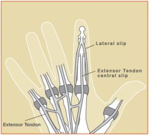
- extensor complex (central slip, lateral bands, and hood) dorsally. These structures attach to and reinforce the joint capsule.
- collateral ligaments on the radial and ulnar sides
- For a dislocation to occur, at least 1, often 2, and sometimes 3 of these structures must be significantly injured.
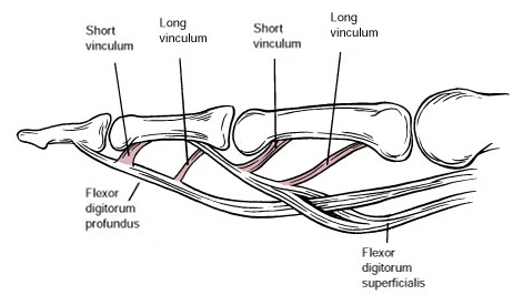
The Flexor Tendons
Radiographic Technique
<a class="external" href="http://www.handandwristinstitute.com/flexor-tendon-injuries/" rel="nofollow" target="_blank">
</a><a class="external" href="http://www.handandwristinstitute.com/flexor-tendon-injuries/" rel="nofollow" target="_blank">http://www.handandwristinstitute.com/flexor-tendon-injuries</a>
The Extensor Tendons
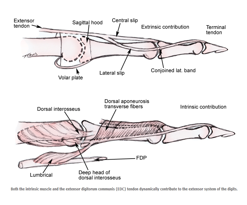
<a class="external" href="http://emedicine.medscape.com/article/1286225-overview" rel="nofollow" target="_blank">http://emedicine.medscape.com/article/1286225-overview</a> 13/2/11The extensor apparatus becomes a complex interplay of intrinsic muscle contribution as it travels from the MP joint distally. Three intrinsic sets of muscles (volar interossei, dorsal interossei, and lumbricals) contribute dynamic function to the extensor system through the lateral slips.
<a class="external" href="http://emedicine.medscape.com/article/1286225-overview" rel="nofollow" target="_blank">http://emedicine.medscape.com/article/1286225-overview</a> 13/2/11
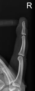
Lateral Finger Technique
Labelling
Labelling
Buddy Taping Splint
Buddy Taping Splint
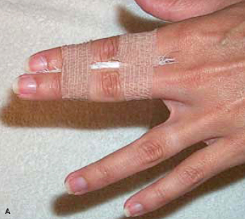
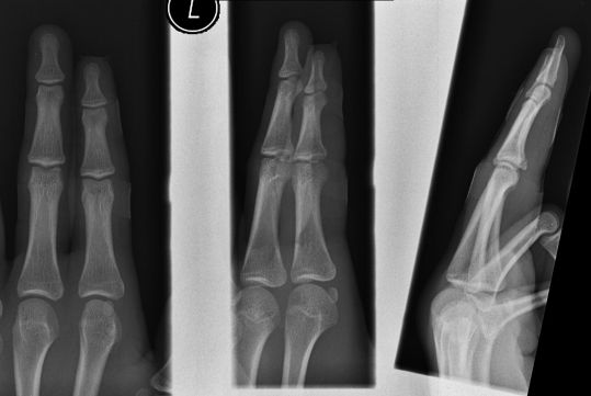
<a class="external" href="http://www.sportmedicine.ru/articles/acute_finger_injuries-part1.pdf" rel="nofollow" target="_blank">JEFFREY C. LEGGIT, CHRISTIAN J. MEKO, Part I. Tendons and Ligaments, Acute Finger Injuries:</a>Patients will present for finger radiography with two fingers taped together. There are a number of radiographic options to consider
- remove the tape splint (don't do this unless the patient routinely removes the splint at home, or you have the express permission of the referring doctor)
- don't perform a lateral view
- perform a lateral view knowing that the anatomy will be partly obscured by the splinting finger
- perform an off-lateral view to ensure some separation of the two fingers
- check your departmental policy re splint removal
This patient presented from fracture clinic with budding taping of the middle and ring fingers. The volar plate fracture is demonstrated on the lateral projection image despite the overlapping finger.
Fixed Flexion Deformity
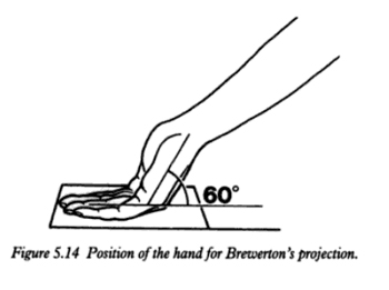
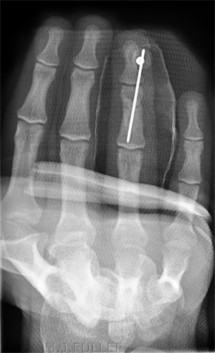
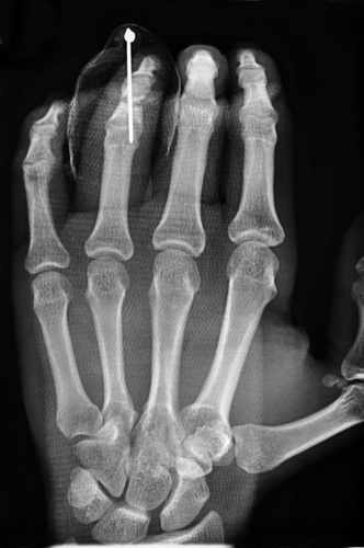
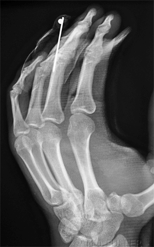
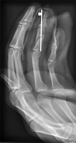
This is the common one- patient will present for follow-up imaging of finger trauma with fixed flexion deformity. This is sometimes a legacy of the splint or caste applied. An exaggerated Brewerton's-like view of the ring finger following K-wire reduction and fixation
Finger or hand radiography
Case Study 1
Case Study 2
Comment
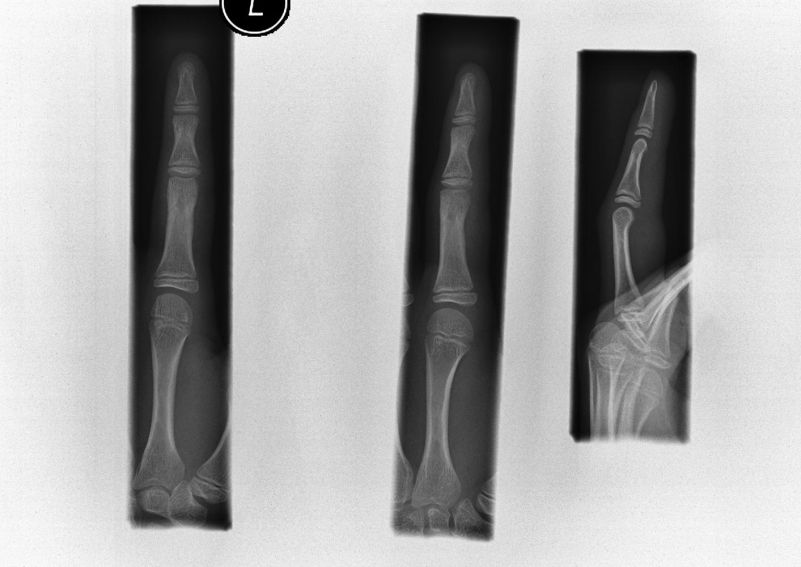
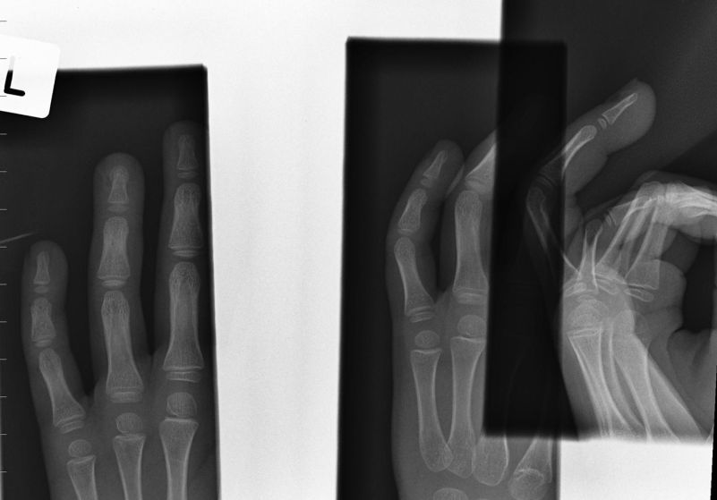
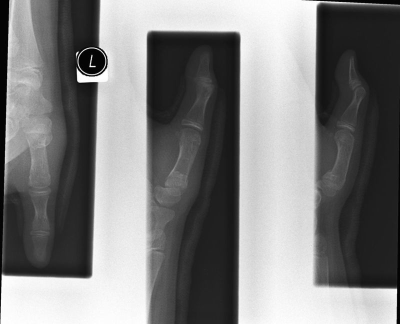
Radiographer 1 has approached this examination with a 'photographic' rather than a 'clinical' focus. Radiographer 1 has performed the routine views for this patient without further thought. Radiographer 2 has taken a clinical approach to this case and asked the question ..."what does the surgeon want from this imaging?" The answer is undistorted and unobstructed views of the fracture. On review of the previous imaging, this was not achieved at the last clinic visit. Radiographer 2 has not only supplied the referring doctor with high quality imaging, he/she has achieved a high level of professional and personal satisfaction.
What Went Wrong?
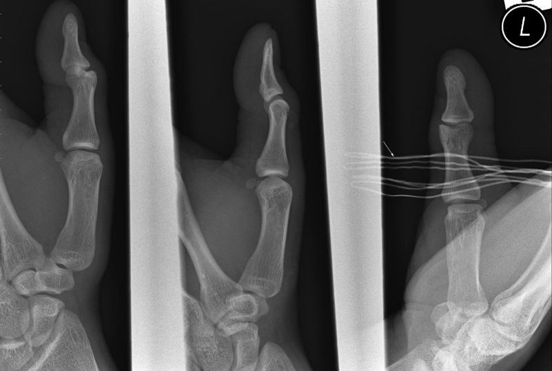
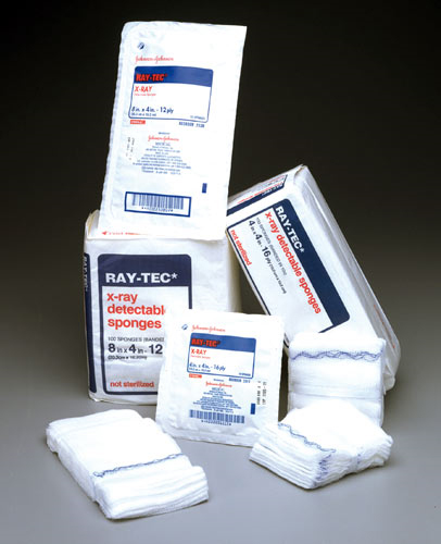
The finger is not labelled- which finger is it? It is also considered good practice to include the adjacent finger/fingers on the PA and/or oblique projections. There is overlap of adjacent images. Raytek gauze (arrowed) overlying thumb on PA projection image. Unless you are familiar with radiopaque sponges, you may not realise that the sponge/gauze has a 'wire' though it.
<a class="external" href="http://www.cardinal.com/us/en/distributedproducts/ASP/JJ2515.asp?cat=med_surg" rel="nofollow" target="_blank">
http://www.cardinal.com/us/en/distributedproducts/ASP/JJ2515.asp?cat=med_surg</a>It can be very easy to forget the orientation of the cassette!
Hyperflexion Injuries
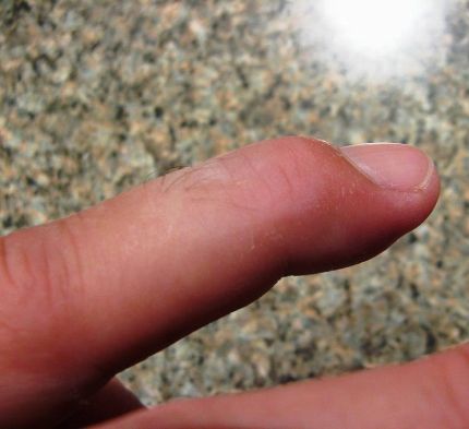
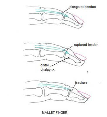
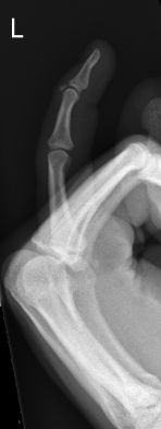
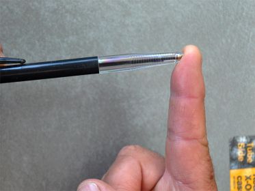
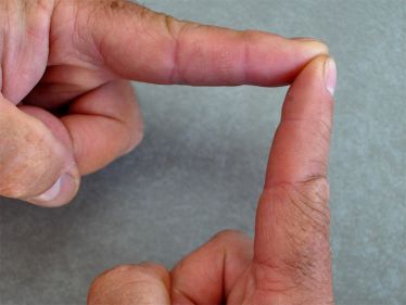
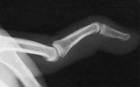
Mallet Finger- DP- lateral slip injury
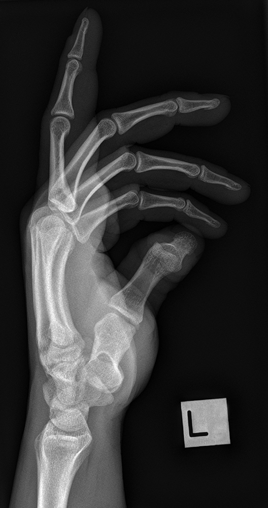
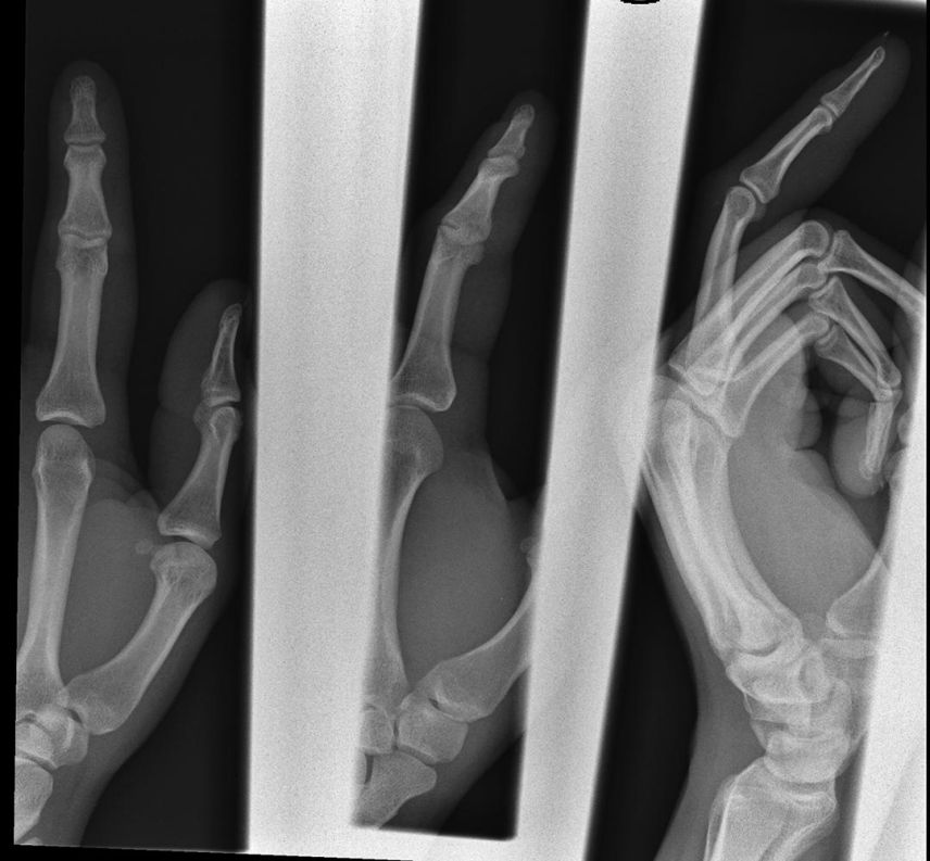
If you use one of these 3 popular lateral finger positioning techniques, you risk masking the extensor tendon injury.
Middle Phalanx Hyperflexion Avulsion Fracture
Volar Plate Injury
Case 1
Case 2
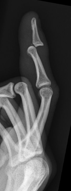
This 62 year old male presented to the Emergency Department after a fall. He was examined and found to have a painful left index finger. He was referred for left index finger radiography. There is a volar plate fracture of the left index finger middle phalanx (arrowed)
Case 3
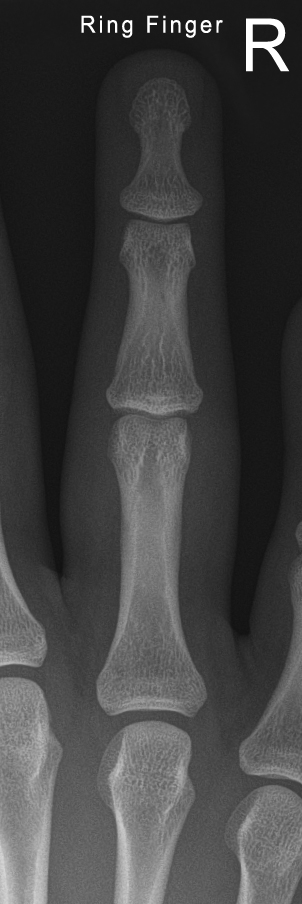
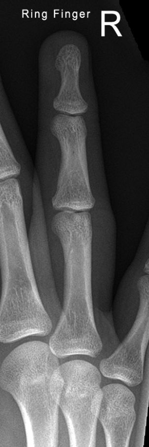
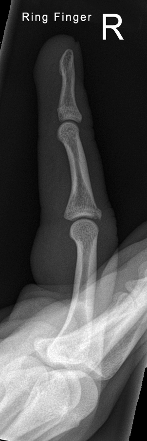
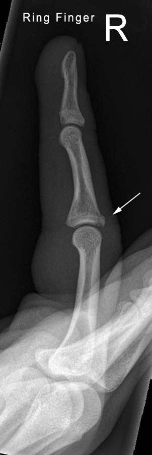
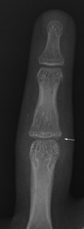
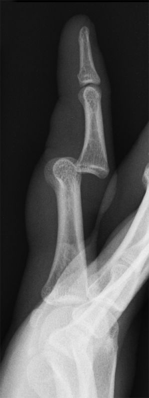
This 12 year old girl presented to the Emergency Department following a fall in which she suffered a hyperextension injury to her left ring finger. She was examined and was found to have a painful swollen proximal IP joint. She was referred for left ring finger radiography. There is a volar plate fracture (arrowed) of the base of the left ring finger middle phalanx best demonstrated on the PA projection image.
Swan neck Deformity
</a>
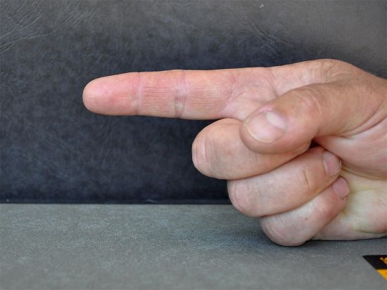
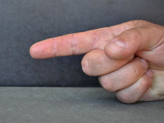
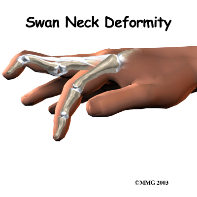
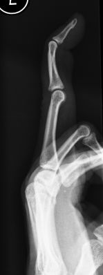
A swan-neck deformity, typically defined as proximal interphalangeal (PIP) joint hyperextension with concurrent distal interphalangeal (DIP) joint flexion, occurs in approximately 50% of patients with RA. However, swan-neck deformity is not unique to RA, because it may also be congenital or traumatic in nature.
<a class="external" href="http://emedicine.medscape.com/article/1244326-overview" rel="nofollow" target="_blank">http://emedicine.medscape.com/article/1244326-overview</a>The swan neck deformity appears similar to the boutonniere deformity.
Boutonniere Deformity
</a>
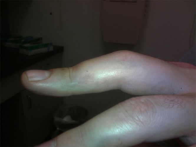
<embed allowfullscreen="true" height="350" src="http://widget.wetpaintserv.us/wiki/wikiradiography/widget/youtubevideo/ccfe2a9ce1cb5473b980aea6d801c220dcbda8d1" type="application/x-shockwave-flash" width="425" wmode="transparent"/> There is only one tendon that is responsible for extending both of the finger joints. This is in contrast to the flexor tendons that are two in number. This extensor tendon divides into three slips (branches). The central one extends the proximal joint and the remaining two (one on each side of the finger) join up and extend the distal joint The boutonniere deformity may not occur right away. It is the imbalance in the extensor hood that results from the torn tendon that eventually causes the deformity. Because the middle phalanx no longer is pulled by the central slip, the flexor tendon on the other side begins to bend the PIP joint without resistance. The lateral bands begin to slide down along the side of the finger where they continue to straighten the DIP joint. Eventually the finger becomes stiff in this position.
<a class="external" href="http://www.eorthopod.com/content/boutonniere-deformity-finger" rel="nofollow" target="_blank">http://www.eorthopod.com/content/boutonniere-deformity-finger</a>This 52 year old lady has developed a boutonniere deformity following an extensor tendon injury. Self explanatory video
Dislocation
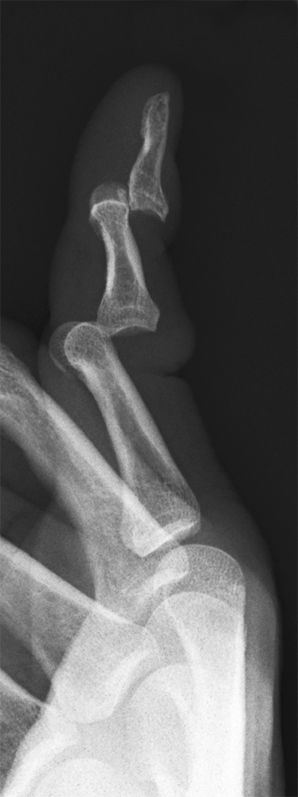
Index finger distal phalax posterior dislocation with volar avulsion fracture. Posterior DIP joint dislocation in a 15 year old male following sports injury. Posterior dislocations of the DIP and PIP joints of the 5th digit in a 63 year old male.
Avulsion fracture of the volar aspect of the distal phalanxPosterior dislocation 2nd MP joint in a 21 year old male following fall.
DP Crush Injury
Labelling
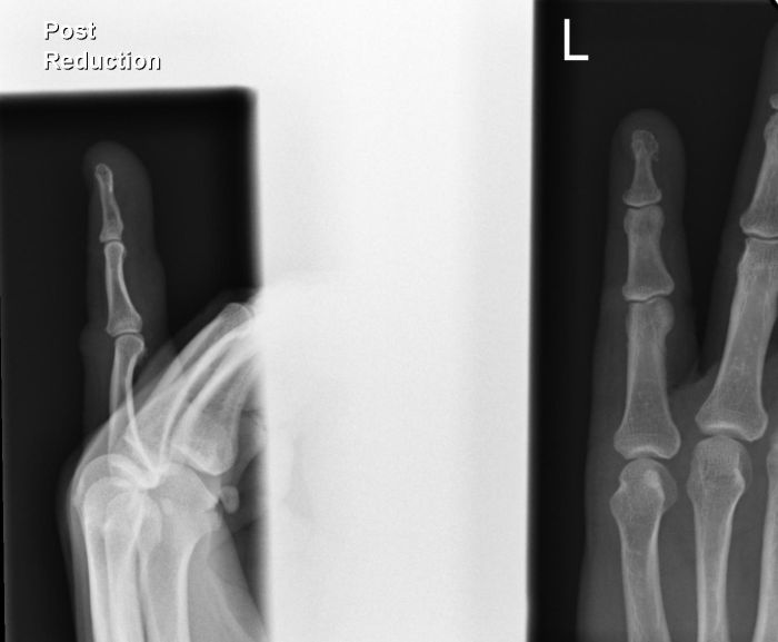
It is good practice to label the image post reduction to indicate that the image is post-treatment. It may also be necessary to label images post reduction 1, post reduction 2 etc where multiple attempts have been made to effect a reduction of the dislocation.
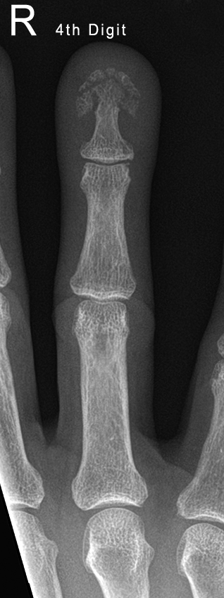
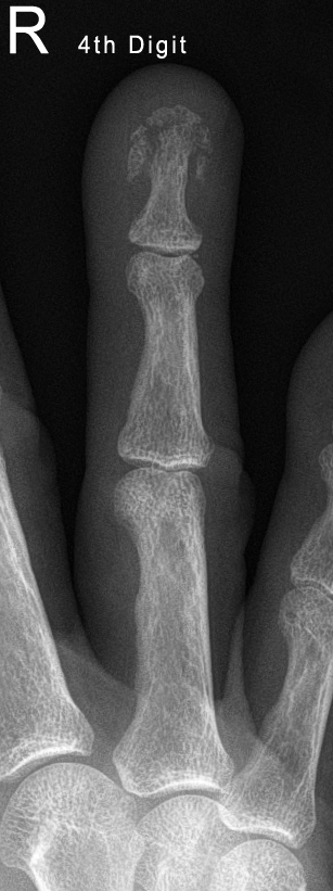
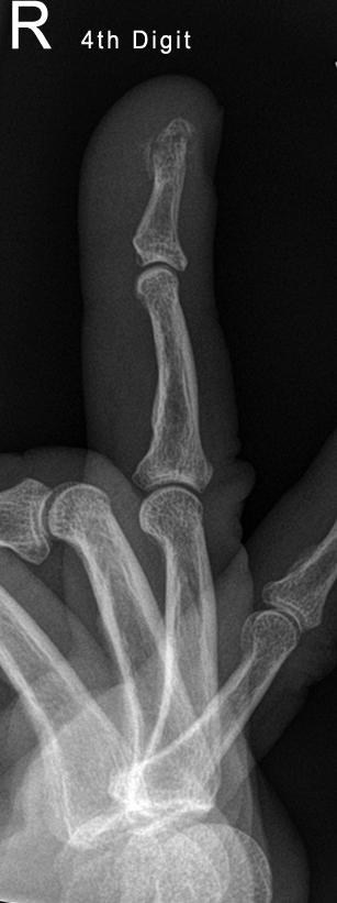
Crush injury in a 61 year old female. There is a comminuted fracture of the terminal tuft of the distal phalanx of the right ring finger. If the fingernail is broken, this is an open fracture- antibiotic therapy is required.
Imaging Amputated Digits
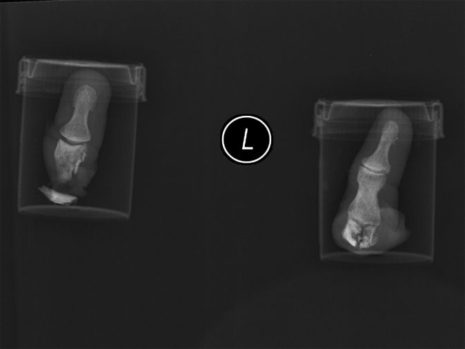
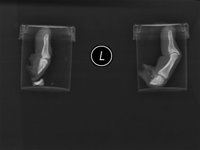
The mechanism of injury resulting in amputated digits is varied. Industrial accidents and home use of circular saws are common mechanisms.
The affected hand and amputated anatomy should all be imaged. These digits have been kept in their specimen containers when imaged. Care should be taken to keep the amputated anatomy clean and moist to maximise chances of successful re-attachment.
The amputated digits should be labelled for side and digit. Whilst it might appear that the identification of the digit is obvious in cases of single digit amputation, confusion with subsequent amputations could occur.
Pathological Fracture
Enchondromas are benign cartilaginous neoplasms that are usually solitary lesions in intramedullary bone. The primary significant factors of enchondromas are related to their complications, most notably pathologic fracture, and a small incidence of malignant transformation, which may be associated with pathologic fracture.
The lesions replace normal bone with mineralized or unmineralized hyaline cartilage, thereby generating a lytic pattern on radiographs or, more commonly, a lytic area containing rings and arcs of chondroid calcifications. The lesions likely arise from cartilaginous rests that are displaced from the growth plate.
<a class="external" href="http://emedicine.medscape.com/article/389224-overview" rel="nofollow" target="_blank">Enchondroma and Enchondromatosis Imaging, Felix S Chew, Catherine Maldjian, MD</a>
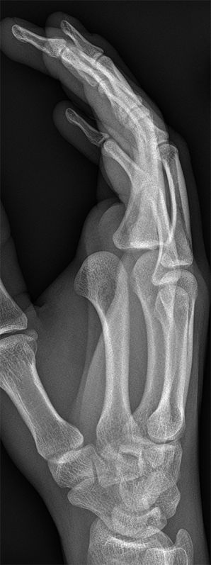
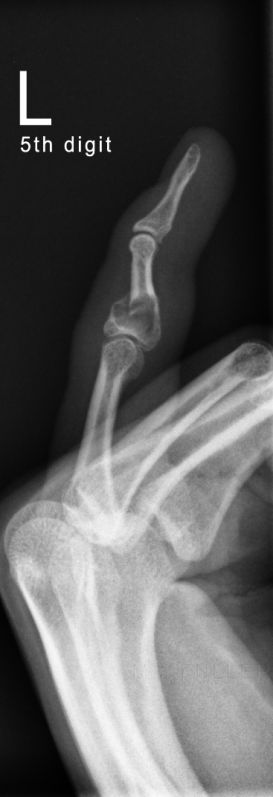
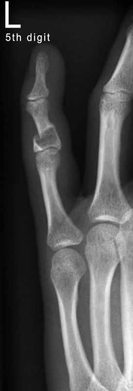
This 32 year old female presented to the ED with finger pain following trivial trauma. There is a sharply marginated lucent lesion sited at the proximal third of the middle phalanx of the 5th digit of the left hand. This is likely to be a enchondroma. There is a transverse pathological frature with a moderate degree of dorsal angulation.
... back to the wikiradiography home page
... back to the Applied Radiography home page
