Fifth Metatarsal Fractures
This page considers all aspects of plain film imaging of metatarsal fractures.
Base of 5th Metatarsal Fractures
Base of 5th metatarsal fractures deserve separate consideration based on their common occurrence and the specific mechanism of injury.
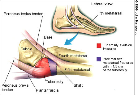
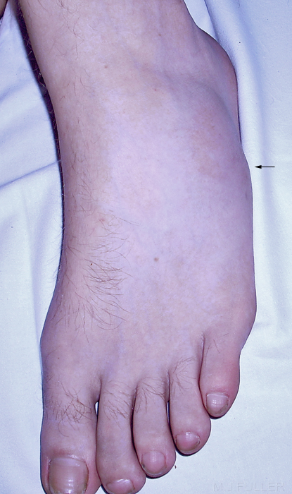
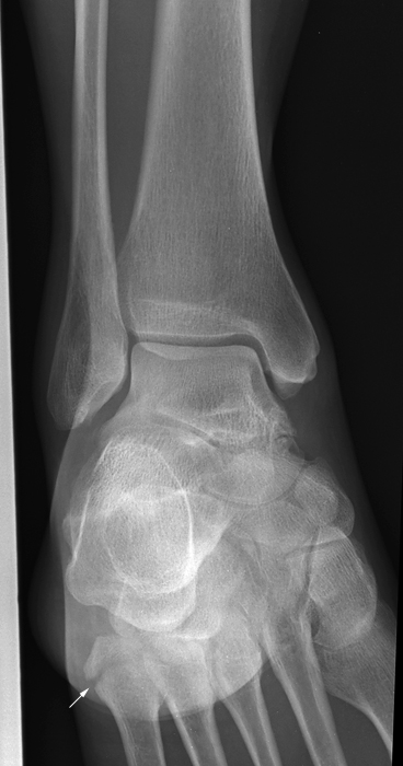
<a class="external" href="http://www.aafp.org/afp/990501ap/2516.html" rel="nofollow" target="_blank">http://www.aafp.org/afp/990501ap/2516.html</a>Fractures of the base of the 5th metatarsal are commonly tuberosity avulsion fractures caused by an inversion/plantar flexion injury of the ankle. The fracture results from an abnormally large tension force transmitted though the peroneus brevis tendon.
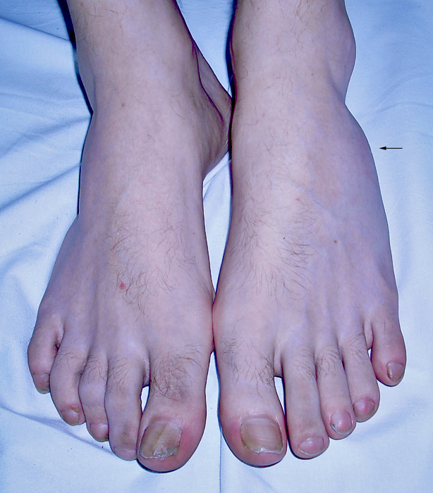
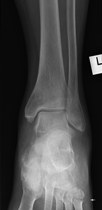
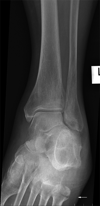
Base of 5th metatarsal fractures present a potential trap for the referring doctor and radiographer alike. The mechanism of injury and other clinical signs may suggest an ankle injury- indeed, there may be considerable pain and swelling associated with the ankle. The referring doctor should pay careful attention to the base of the 5th metatarsal when examining all patients presenting with a history of ankle inversion injury. Similarly, the radiographer should also consider the possibility clinically and radiographically that the patient who is referred for ankle radiography following an inversion injury may have a base of 5th metatarsal avulsion fracture.
Mechansim of Injury
<embed allowfullscreen="true" height="350" src="http://widget.wetpaintserv.us/wiki/wikiradiography/widget/youtubevideo/1fca3df9d5b45580404685feb5d34e827bc9e1f1" type="application/x-shockwave-flash" width="425" wmode="transparent"/> <embed allowfullscreen="true" height="350" src="http://widget.wetpaintserv.us/wiki/wikiradiography/widget/youtubevideo/272a5dc1e337888220b5b3de7af961d89f22075d" type="application/x-shockwave-flash" width="425" wmode="transparent"/> <embed allowfullscreen="true" height="350" src="http://widget.wetpaintserv.us/wiki/wikiradiography/widget/youtubevideo/f2127ae1b4750afa4c44f79ff55e96a3d6169be3" type="application/x-shockwave-flash" width="425" wmode="transparent"/> <embed allowfullscreen="true" height="350" src="http://widget.wetpaintserv.us/wiki/wikiradiography/widget/youtubevideo/76baad22a4e6784df4cc5508c81d7dbfcc4cfcaf" type="application/x-shockwave-flash" width="425" wmode="transparent"/>
Fracture, Apophysis or Both?
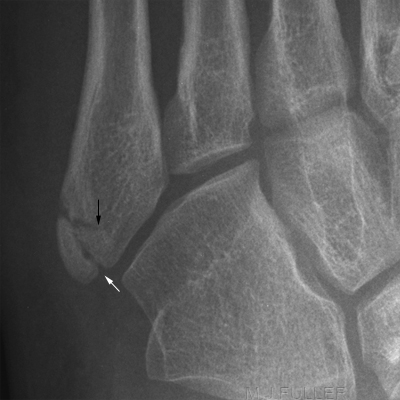
The development of a secondary center of ossification (apophysis) at the base of the fifth metatarsal can be mistaken radiographically as a fracture site. In girls nine to 11 years of age and boys 11 to 14 years of age, the apophysis becomes visible on plain radiographs as a fleck of calcification adjacent to the fifth metatarsal shaft. The apophysis has an oblique orientation, with the radiolucency aligned parallel to the fifth metatarsal diaphysis.
<a class="external" href="http://www.aafp.org/afp/990501ap/2516.html" rel="nofollow" target="_blank">SCOTT M. STRAYER, CPT, USAF, MC, STEVEN G. REECE, M.D., and MICHAEL J. PETRIZZI, M.D.
</a><a class="external" href="http://www.aafp.org/afp/990501ap/2516.html" rel="nofollow" target="_blank">
</a> <a class="external" href="http://www.aafp.org/afp/990501ap/2516.html" rel="nofollow" target="_blank"> Fractures of the Proximal Fifth Metatarsal
http://www.aafp.org/afp/990501ap/2516.html</a>
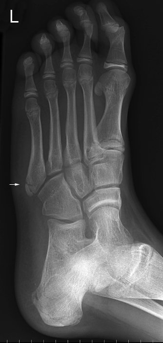
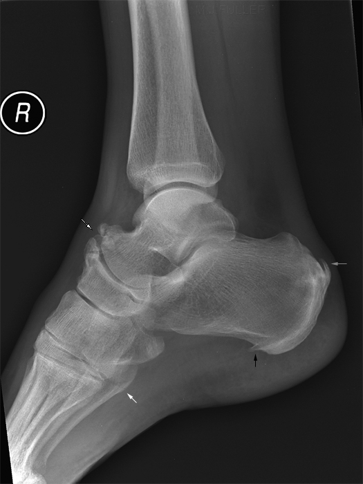
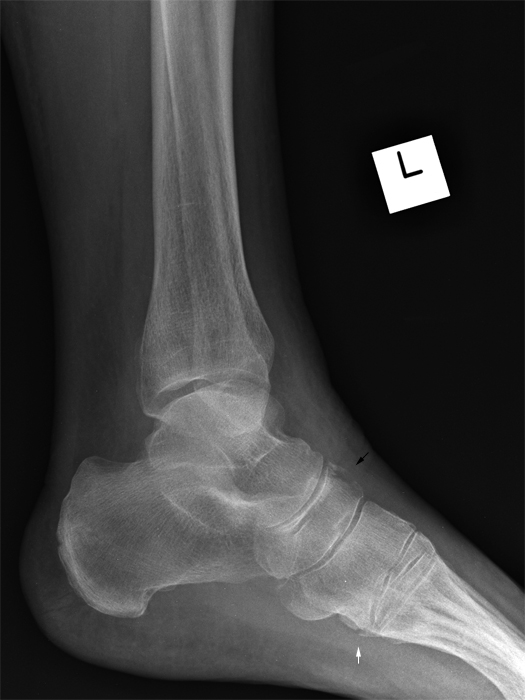
This 13 year old boy presented to the Emergency Department after an inversion injury to his left ankle while playing on a trampoline. His oblique foot image demonstrates 2 lucent lines resembling fractures at the base of the 5th metatarsal (arrowed). Are they fractures, apophysis or both?The white arrow identifies the normal apophysis. The characteristic features are the orientation of the lucent line (longitudinal oblique rather than transverse) and the rounded corners and sclerotic margins. The black arrow is pointing to the avulsion fracture.
Clinical Presentation
Quality of Referral
Referrals for ankle and foot radiography are often confused. Patients who have clear clinical signs of a base of 5th metatarsal fracture (mechanism + localised swelling + localised pain) will sometimes be referred, in error, for ankle radiography. Many referrers simply ask for ankle and foot radiography for all injuries involving ankle and/or foot. This is an area where the radiographer must employ a patient-centred rather than a picture-centred approach to radiography- ask the patient about their mechanism of injury, examine the patient, and if there are any discrepancies between the patient's presentation and the requested imaging communicate your concerns with the referring doctor. Where your scope of practice allows for modification to the requested views/anatomy, modify your technique to suit.
Jones Fracture Classification Confusion
Fifth Metatarsal
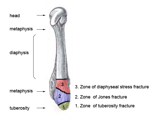
adapted from<a class="external" href="http://thefootblog.wordpress.com/2006/11/07/jones-fracture-a-case-report-with-special-emphasis-on-fixation-placement/" rel="nofollow" target="_blank">http://thefootblog.wordpress.com/2006/11/07/jones-fracture-a-case-report-with-special-emphasis-on-fixation-placement</a>The Jones fracture describes a fracture to the base of the fifth metatarsal. It was named after Sir Robert Jones, who in 1902, sustained this fracture while dancing around a maypole at a garden dance. The injury is caused by stress plantarflexion and inversion. <a class="external" href="http://thefootblog.wordpress.com/2006/11/07/jones-fracture-a-case-report-with-special-emphasis-on-fixation-placement/" rel="nofollow" target="_blank">(http://thefootblog.wordpress.com/2006/11/07/jones-fracture-a-case-report-with-special-emphasis-on-fixation-placement)</a>
There has been some debate and confusion as to the correct descriptions of fractures of the proximal fifth metatarsal. Al Kline notes the followingIn reviewing the literature, these ‘zones’ differ and some are delineated by distance from the end of the bone. For instance, fractures that are within 1.5 cm to the end of the styloid process are considered an ‘avulsion’ fracture. Anything distal to that is a ‘metatarsal’ fracture.
There has also been 3 zones of fracture descrbed in metatarsal base fractures:1) zone of tuberosity fracture
2) zone of Jones fracture and
3) zone of diaphyseal stress fracture.
For our simplified classification, it is fractures that involve the styloid process that are considered true ‘avulsion’ fractures. Fractures to the body of the fifth metatarsal base are considered ‘base fractures’ , and fractures distal to the body of the fifth metatarsal base are considered ‘metatarsal’ or diaphyseal fractures. In most of the literature, this is also the most common region for stress fractures associated with a Jones fracture.
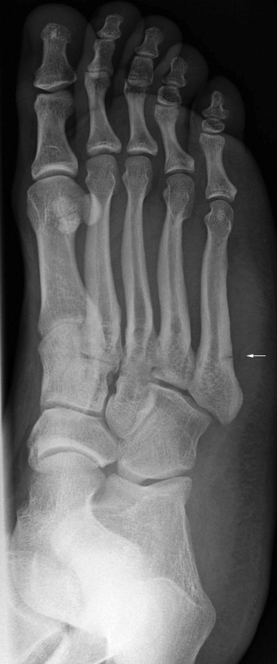
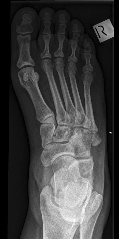
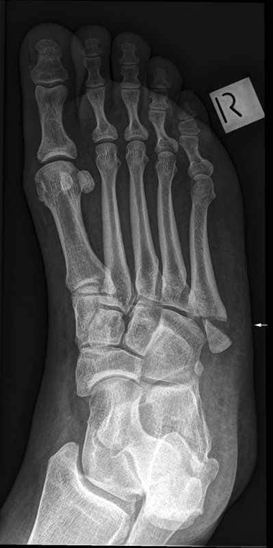
Many Radiologists prefer to apply the eponym rule - that is, dont use eponyms...simply describe what you see.Jones fracture of the proximal 5th metatarsal (white arrow)
Case 1
Case 2
Case 3
When Should the Base of the 5th Metatarsal be Included with Ankle Radiography?
The short answer is the base of the 5th metatarsal should be included in ankle radiography whenever there is a reasonable chance of 5th metatarsal bony pathology based on consideration of mechanism of injury and clinical signs. The inclusion of the base of the 5th metatarsal on plain film ankle imaging is arguably not required when foot radiography is to be performed along with ankle radiography.
The following should be taken into consideration
- departmental protocols (some departments require the base of the 5th metatarsal to be included with all ankle radiography- some consider this simply good practice for all ankle radiography)
- mechanism of injury- did the patient present following an ankle inversion injury?
- Are you performing foot radiography as well as ankle radiography?
- Is there pain or swelling over the 5th metatarsal?
You could argue that base of 5th metatarsal fractures should be considered and treated as all other fractures- clinical exam followed by appropriate imaging (why include the base of the 5th metatarsal when it is not clinically fractured?). The difficulty with this view is that errors are made- the clinical signs of base of 5th metatarsal fracture are not always picked up by the referring doctor. Another important consideration is that most patients with base of 5th metatarsal fractures will be exquisitely tender in this region- unfortunatley some are not, possible due to distracting ankle pain... beware.
