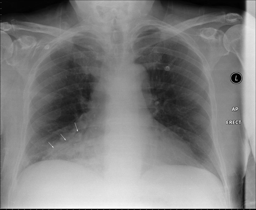
False Consolidation
Jump to navigation
Jump to search
Introduction
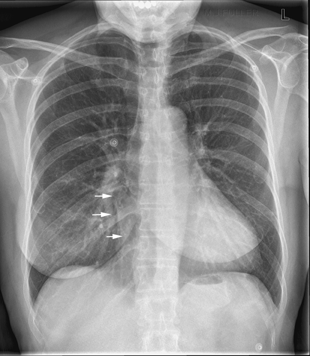
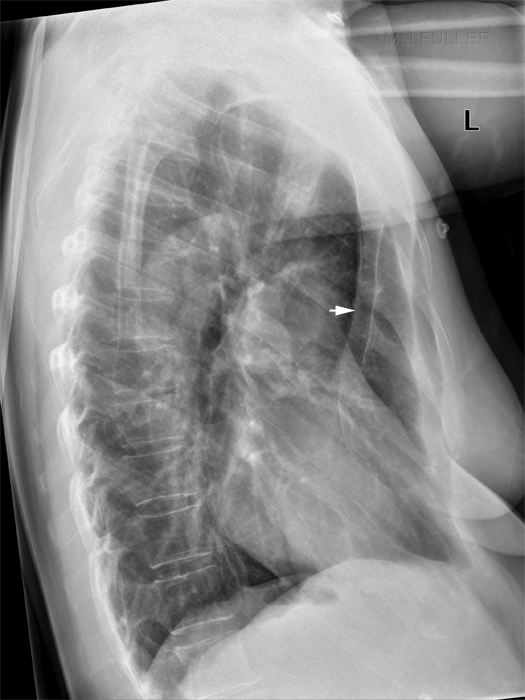
False RML Collapse/Consolidation in Patients with Pectus Excavatum
False RML Collapse/Consolidation in Patients with Pericardial fat
... back to the Applied Radiography home page
Identification of consolidation on chest images is a useful skill for radiographers. Care must be taken to avoid declaring every lung opacity a consolidation. There are a variety of causes of lung opacity and a number of known false signs. This page looks at some of the other consolidation-like appearances.
False RML Collapse/Consolidation in Patients with Pectus Excavatum
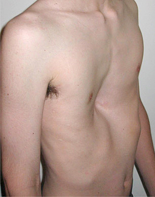
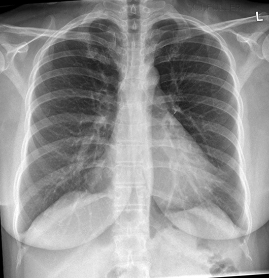
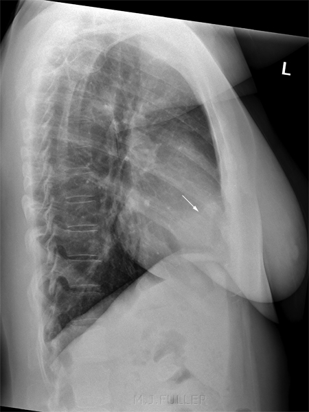
This patient presented for chest radiography with a history of recent facial nerve palsy. There is loss of the right cardiac border. There is no abnormal RML opacity. There is evidence of pectus excavatum (depression of the sternum). Pectus excavatum is a known cause of false RML disease and is likely to be a contributing cause of the pseudo-silhouette sign on the PA image. In these patients the heart tends to be displaced towards the left as a result of the limited space between the depressed sternum and the spine.Other contributing factors could be pericardial fat (although none is seen in this patient) and overlying pulmonary vessel. Pectus Excavatum
<a class="external" href="http://en.wikipedia.org/wiki/File:Pectus1.jpg" rel="nofollow" target="_blank">http://en.wikipedia.org/wiki/File:Pectus1.jpg</a>
False RML Collapse/Consolidation in Patients with Pericardial fat
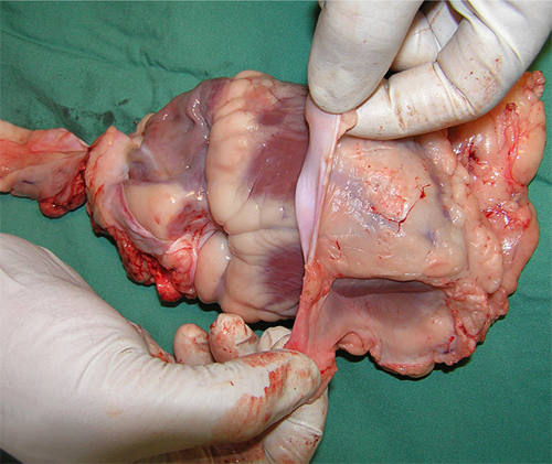
Pericardial Fat
<a class="external" href="http://jcmr-online.com/content/11/1/15" rel="nofollow" target="_blank">Validation of cardiovascular magnetic resonance assessment of pericardial adipose tissue volume</a><a class="external" href="http://jcmr-online.com/content/11/1/15" rel="nofollow" target="_blank">Adam J Nelson , Matthew I Worthley , Peter J Psaltis , Angelo Carbone , Benjamin K Dundon , Rae F Duncan , Cynthia Piantadosi , Dennis H Lau , Prashanthan Sanders , Gary A Wittert and Stephen G Worthley</a><a class="external" href="http://jcmr-online.com/content/11/1/15" rel="nofollow" target="_blank">Cardiovascular Research Centre, Royal Adelaide Hospital & Disciplines of Medicine and Physiology, University of Adelaide, Adelaide, SA, Australia</a>This is a sheep's heart with the pericardium partly dissected off. Note the pericardial fat.
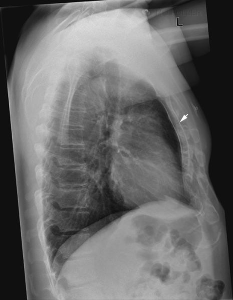
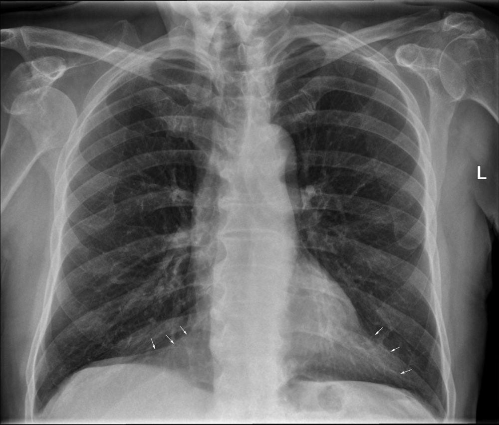
Felson (<a class="external" href="http://www.amazon.com/Chest-Roentgenology-Benjamin-Felson/dp/0721635911/ref=sr_1_2?ie=UTF8&s=books&qid=1252240078&sr=1-2" rel="nofollow" target="_blank">Chest Roentgenology, W.B. Saunders, 1973, p55</a>) notes that "For reasons that escape me, fat in the thorax is often impossible to differentiate from water density. Perhaps the adjacent pulmonary gas density has something to do with this illusion." This is an interesting point given the clear differentiaton between fat and water density structures seen in the abdominal plain film.
Case 1
Case 2
... back to the Applied Radiography home page
