Elbow/Forearm

From wikiRadiography
Jump to navigation Jump to search

Elbow Surface Anatomy


Other pages of interest

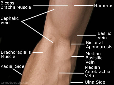
Elbow Anterior surface:


Elbow/Forearm - wikiRadiography

Three bones form the elbow joint, the humerus of the upper arm, and the paired radius and ulna of the forearm.

The elbow joint is a hinge joint and can perform flexion and extension.

The complex action of turning the forearm over (pronation or supination) happens at the articulation between the radius and the ulna (this movement also occurs at the wrist joint).

In the anatomical position with the forearm supinated the radius and ulna lie parallel to each other. During pronation, the ulna remains fixed, and the radius rolls around it at both the wrist and the elbow joints. In the pronated position, the radius and ulna appear crossed.

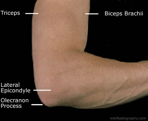
Elbow Posterior Surface

Elbow/Forearm - wikiRadiography

On the humerous you are able to feel the medial and lateral epicondyles. The olecranon process is the large bony protuberance on the posterior of the elbow joint. This is referred to in laymans terms as the "funny bone".


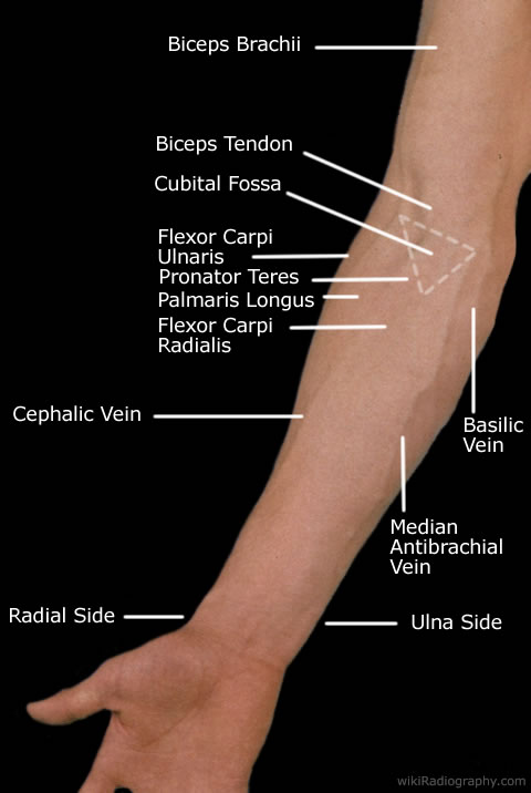
Forearm Anterior Surface:


Elbow/Forearm - wikiRadiography The forearm contains two long bones, the radius and the ulna. At the proximal end they articulate with the humerus forming the elbow joint. At the distal end they articulate with the carpal bones forming the wrist joint.

Connecting inbetween the radius and ulna is the interosseous membrane (fibrous sheet). The interosseus membrane divides the forearm into anterior and posterior compartments, serves as a site of attachment for muscles of the forearm, and transfers forces from the radius, to the ulna and to the humerus.

The anterior surface of the forearm usually has less hair than the posterior surface.

The radial and ulnar arteries, and their branches, supply the blood to the forearm. These usually run on the anterior face of the radius and ulna down the whole forearm.

The main superficial veins of the forearm are the cephalic, median antebrachial and the basilic vein. These veins can be used for cannularisation or venipuncture, although the cubital fossa is a preferred site for getting blood.

In human anatomy, the biceps brachii is a muscle located on the humerus. The biceps has several functions, the most important simply being to flex the elbow and to rotate the forearm. Its insertion is on the radial tuberosity and its tendon can be felt as it crosses the elbow joint. The biceps brachii is one of the best known muscle, as it lies fairly superficially, and is often well-defined even in non-athletes.

The superficial muscles in the anterior of the forearm serve to primarily flex the wrist and fingers. These muscles from lateral to medial are the , flexor carpi ulnaris, palmaris longus, flexor carpi radialis and pronator teres.

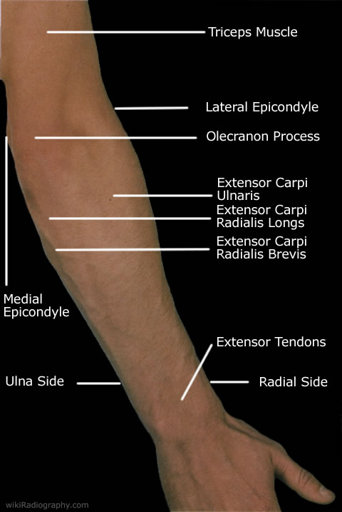
Forearm Posterior Surface:


Elbow/Forearm - wikiRadiography The bones that make up the elbow joint are most easily palpated posteriorly.

The superficial muscles in the posterior of the forearm serve to primarily extend the wrist and fingers. The primary superficial muscles are the extensor carpi radialis longus, extensor carpi radialis brevis and the extensor carpi ulnaris.

The tendons that attach to the extensor muscles run along the posterior of the forearm towards the distal end. These are able to be felt sliding under the surface if you flex and extend your fingers continously.

Distally the head of the ulna is able to be palpated and the dorsal tubercle on the radial side.

The posterior skin surface of the forearm has more hair than the anterior.


Go Back to Surface Anatomy Index