Digital Tomosynthesis
Jump to navigation
Jump to search
Digital tomosynthesis is a medical imaging technique using a flat panel detector and tube rotation producing a series of slices at different depths. It provides better visualization than projection radiography.
Digital tomosynthesis is analogous to geometric tomography. The difference is that in digital tomosynthesis, the x-ray tube makes one sweep and produces many slices. While with geometric tomography, each sweep produces one slice. This is enabled by the rapid readout capabilities of flat panel digital detectors. A series of low dose exposures are made during the sweep which are reconstructed to produce slices at different planes within the patient. [1, 2]
Most developments in tomosynthesis have been in breast imaging, but orthopedic, chest, and urinary applications have also been used. [2]
Angiography
The dose in chest tomosynthesis is about 0.12 mSv, 3 times more than digital radiography but three times less than chest CT. [2]
Parallel Path
The tube moves in a plane parallel to the detector plane and the detector may also move in its plane. Typically used in chest and abdominal. Simplest reconstruction algorithm, maintains uniform magnification. [4]
Full Isocentric Motion
The tube and detector are fixed rigidly with respect to eachother and move in tandem in a circular path around the patient. Used in cone-beam CT, in c-arm imagers and radiation oncology to verify patient position. Can provide excellent reconstructions but with a more complicated algorithm. [4]
Partial Isocentric Motion
The detector remains stationary, x-ray tube moves in an arc above the detector, used in breast tomosynthesis because of the ease of constructing a compact rotational gantry for the x-ray tube. Leads to variable magnification and different tube orientations and can therefore distort small structures unless care is taken in the back-projection process. [4]
Conventional filtered back projection cannot be used because it requires a complete data set. [3]. Instead, shift-and-add reconstruction is used.
In addition to SAA, other more sophisticated mathematical de-blurring techniques can be used to improve image quality and slice. [1]
1. Bushberg JT, Siebert JA, Leidholdt EM, Boone JM. The essential physics of medical imaging. 2nd ed. New York, NY: Lippincott Williams & Wilkins; 2002.
2. Brice J. Will new DR technologies offer alternative to CT? Part 2. <a class="external" href="http://www.auntminnie.com/index.aspx?Sec=sup&Sub=xra&Pag=dis&ItemId=90041" rel="nofollow" target="_blank">http://www.auntminnie.com/index.aspx?Sec=sup&Sub=xra&Pag=dis&ItemId=90041</a>. Updated March 25, 2010. Accessed October 28, 2010.
3. X-ray computed tomography. http://www.wikipedia.org/wiki/X-ray_computed_tomography. Updated October 21, 2010. Accessed October 28, 2010.
4. Dobbins JT. Tomosynthesis imaging: At a translational crossroads. Medical Physics. 2009;36(6):1956-1967.
5. Proposals:Orientation. Retrieved from http://www.vtk.org/Wiki/Proposals:Orientation. Updated June 2, 2010. Accessed February 4, 2011.
Digital tomosynthesis is analogous to geometric tomography. The difference is that in digital tomosynthesis, the x-ray tube makes one sweep and produces many slices. While with geometric tomography, each sweep produces one slice. This is enabled by the rapid readout capabilities of flat panel digital detectors. A series of low dose exposures are made during the sweep which are reconstructed to produce slices at different planes within the patient. [1, 2]
Most developments in tomosynthesis have been in breast imaging, but orthopedic, chest, and urinary applications have also been used. [2]
Diagnostic Uses
Breast
Bone
IVP
Chest
Angiography
Dental
Advantages and Disadvantages
Advantages
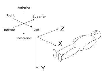
3D plane orientation. [5] |
- Tomosynthesis can provide resolution of 0.22-0.4mm in the x- and y- planes. In the z-plane the resolution is 5mm for chest and 1mm for breast.
- Higher resolution in X- and y-planes than CT. [4]
- Dose-efficient compared to geometric tomography [1]
Disadvantages
- Incomplete data set, therefore thinner slices than CT in the z-plane. [4]
- The resultant data set does not consist of isotropic voxels.[4]
Dose
The dose in chest tomosynthesis is about 0.12 mSv, 3 times more than digital radiography but three times less than chest CT. [2]
Process
The x-ray tube follows a small rotation angle of < 40°[4]. During the sweep a small number of discrete exposure are made (for example, 10 exposures). [3]Geometry of Motion
Parallel Path
The tube moves in a plane parallel to the detector plane and the detector may also move in its plane. Typically used in chest and abdominal. Simplest reconstruction algorithm, maintains uniform magnification. [4]
Full Isocentric Motion
The tube and detector are fixed rigidly with respect to eachother and move in tandem in a circular path around the patient. Used in cone-beam CT, in c-arm imagers and radiation oncology to verify patient position. Can provide excellent reconstructions but with a more complicated algorithm. [4]
Partial Isocentric Motion
The detector remains stationary, x-ray tube moves in an arc above the detector, used in breast tomosynthesis because of the ease of constructing a compact rotational gantry for the x-ray tube. Leads to variable magnification and different tube orientations and can therefore distort small structures unless care is taken in the back-projection process. [4]
Reconstruction
Conventional filtered back projection cannot be used because it requires a complete data set. [3]. Instead, shift-and-add reconstruction is used.
- Sometimes called shift-and-sum
- Filtered back projection used to reconstruct slices
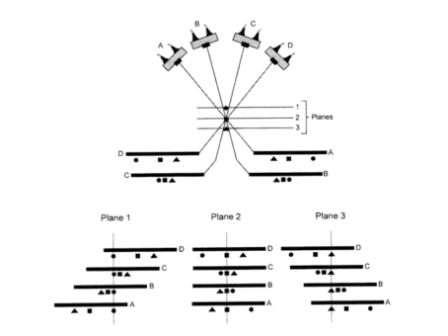
- Digital images are shifted and added to produce images
- Projections of the same point in the patient are superimposed and the images are added
- All planes of interest in the acquisition volume can be reconstructed, using different shift distance
- With no shift distance, images are aligned and plane 2 is in focus, which is coincident with the focal plane of the scan, plane 2 is a digital example of geometric tomography. [1]
In addition to SAA, other more sophisticated mathematical de-blurring techniques can be used to improve image quality and slice. [1]
References
1. Bushberg JT, Siebert JA, Leidholdt EM, Boone JM. The essential physics of medical imaging. 2nd ed. New York, NY: Lippincott Williams & Wilkins; 2002.
2. Brice J. Will new DR technologies offer alternative to CT? Part 2. <a class="external" href="http://www.auntminnie.com/index.aspx?Sec=sup&Sub=xra&Pag=dis&ItemId=90041" rel="nofollow" target="_blank">http://www.auntminnie.com/index.aspx?Sec=sup&Sub=xra&Pag=dis&ItemId=90041</a>. Updated March 25, 2010. Accessed October 28, 2010.
3. X-ray computed tomography. http://www.wikipedia.org/wiki/X-ray_computed_tomography. Updated October 21, 2010. Accessed October 28, 2010.
4. Dobbins JT. Tomosynthesis imaging: At a translational crossroads. Medical Physics. 2009;36(6):1956-1967.
5. Proposals:Orientation. Retrieved from http://www.vtk.org/Wiki/Proposals:Orientation. Updated June 2, 2010. Accessed February 4, 2011.

