Digital Double Dipping in Chest Trauma Radiography
Digital double dipping refers to the practice of extracting two images from a single exposure by post-processing alterations and saving as two separate images. I have argued the case that there are occasions where digital double dipping can result in an undesirable compromise. I am arguing the case on this page that digital double dipping can result in a significant benefit in the resuscitation room. The two cases are patients who have been involved in motor-vehicle accidents prior to presenting to the resus room.
Case 1
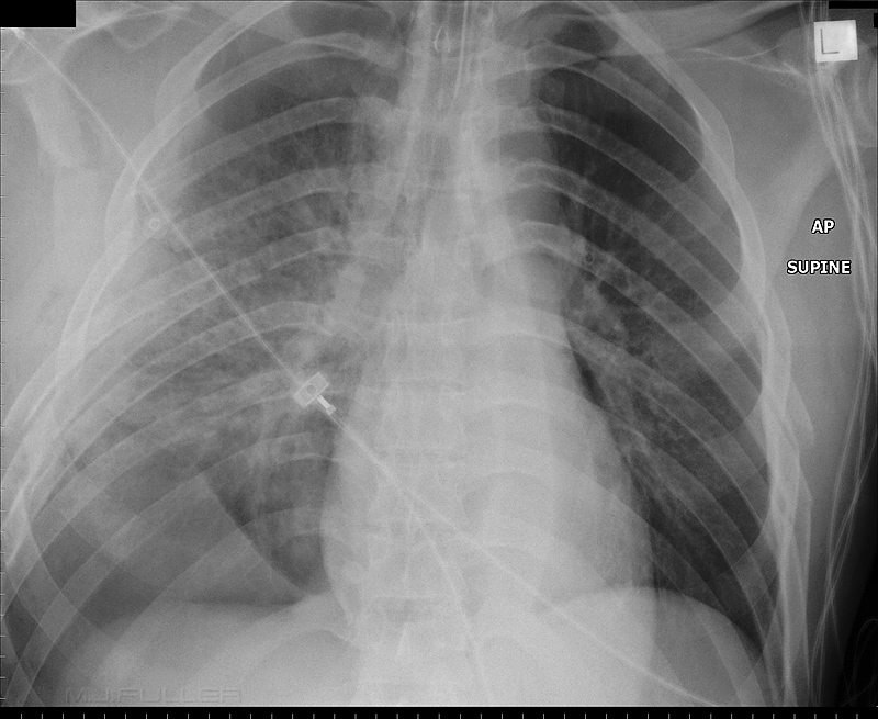
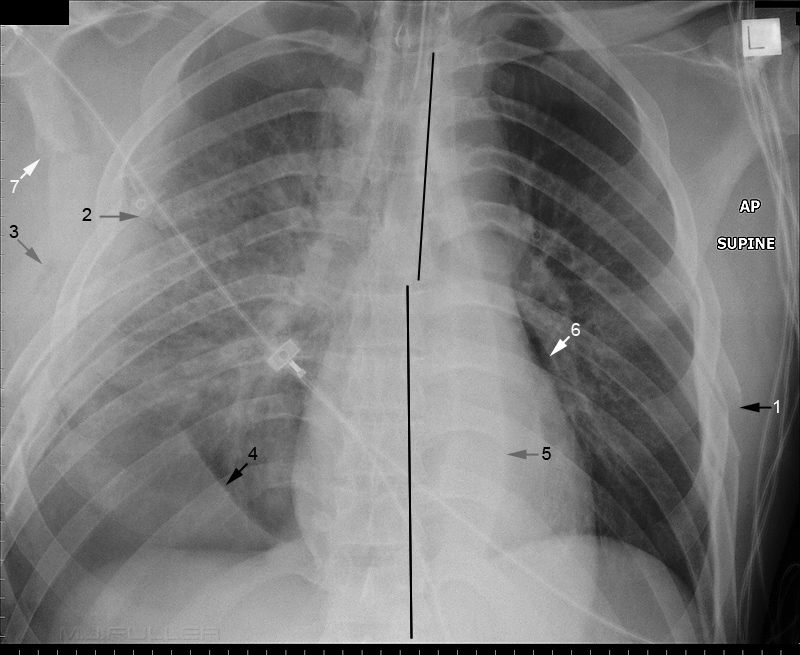
This patient presented to the resus room following a motorvehicle accident. A supine chest image is shown below. What are the findings? Hint- there are more than you think... I found 10!
 | |
 | |
| 1. There are rib fractures on the left 2. There are multiple rib fractures on the right with the suggestion of a flail segment 3. There is subcutaneous emphysema on the right 4. This line suggests a haemopneumothorax. The pleural air has tracked to the least dependent point in the chest and is sharply contrasted against the blood in the pleural space. (see page on supine pneumothorax) 5. There is extreme widening/prominence of the left paraspinal line 6. There is air in the pleural space indicating pneumothorax 7. There is a fractured scapula 8. There is a marked change in the alignment of the thoracic pedicles at the level of T7/8 indicating a displaced thoracic spine fracture 9. There is increased airspace opacity in the right lung suggestive of pulmonary contusion (not marked) 10. There is a suggestion of "etched diaphragm" on the left indicating pneumothorax. (not marked) | |
Case 2
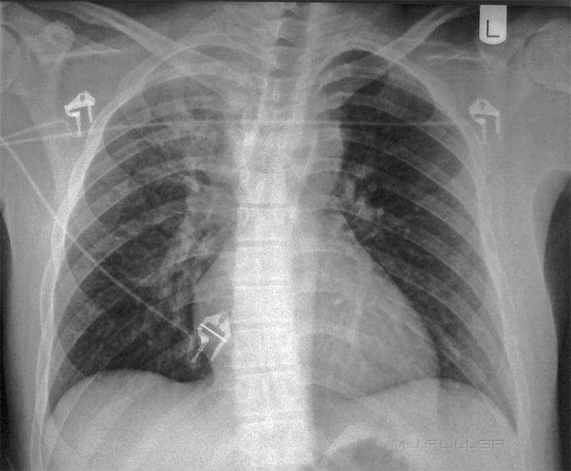
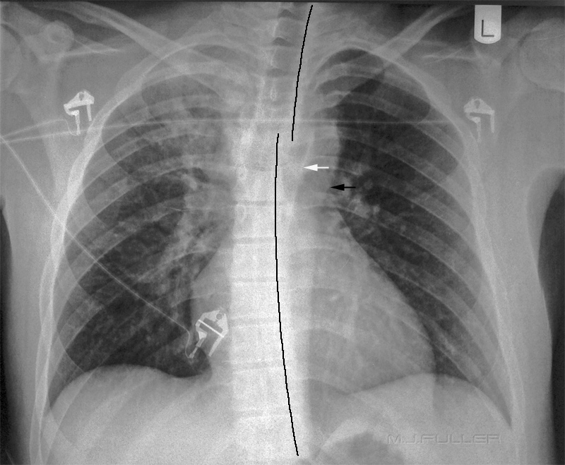
This patient similarly presented to the resus room following a motorvehicle accident. A supine chest image is shown below. What are the findings? Hint- not so many this time.
 | |
Discussion
Radiography in the resus room is different to radiography elsewhere. In the resus room information is needed quickly and there are reasonable grounds for considering a different balance to the speed/quality/dose equation to achieve timely information. The two cases above would have benefited enormously from an exposure that was sufficient to demonstrate the thoracic spine clearly. The exposure technique used was to use the Automatic Exposure Device with the side chamber(s). The diagnosis of the thoracic spine injuries would have been facilitated if the central chamber was used or if an equivalent manual exposure was employed. You could argue that the injuries would be picked up on a subsequent chest CT scan but it should be considered that you are moving a patient with a serious thoracic spine injury onto the CT table and you don't know it!. The radiographer attempted to post-process the chest image in case 2 to produce ap AP thoracic spine image- this largely failed because of lack of exposure.
I have heard it suggested that the automatic exposure device using central chamber should be used for all PA/AP chest radiography with digital systems on the grounds that you can demonstrate pathology that might be otherwise be obscured by the heart and diaphragms. There has never been a better case for the application of that thinking than in these cases of severe trauma to the chest .
back to the Applied Radiography home page
