Conventions, Customs, Cultures, Common Practices and Quirks in Radiography
How do You spell the word X-ray?There are many practices in radiography that have an unclear status and an unclear acceptance. This page considers practices and conventions in radiography . If you have never heard of them, you might consider them idiosyncrasy rather than good practice. Others you may consider to have had their day and dismiss them as anachronisms. If you would like to add your comments on these conventions, customs etc, please start a "thread"at the bottom of this page.
This is not an attempt at humour. The word "X-ray" can be spelt with a lower case "x" or an upper case "X". Originally, the “X” in "X-ray" simply stood for unknown. But is that the case today? A distinction can be made between "X-ray" and terms like "e-mail" where the "e" is an abbreviation and terms like "S-bend" where the "S" describes a shape. For what it's worth, "X-ray" appears to have greater acceptance and greater usage. Lower case "x-ray" spelling is, I believe, an acceptable spelling, and "xray" is simply wrong.
Viewing X-ray Images
The Anatomical Position
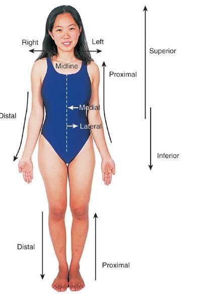
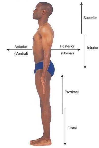
There are a few practices (and exceptions to practices) that relate to viewing X-ray images. The first amongst these is the convention of viewing X-ray images as if you were looking at the patient standing in the anatomical position as shown below.
<a class="external" href="http://nursingassistants.net/2008/02/04/spot-light-medicalnursing-jargon/" rel="nofollow" target="_blank">http://nursingassistants.net/2008/02/04/spot-light-medicalnursing-jargon/</a>
<a class="external" href="http://nursingassistants.net/2008/02/04/spot-light-medicalnursing-jargon/" rel="nofollow" target="_blank">http://nursingassistants.net/2008/02/04/spot-light-medicalnursing-jargon/</a>The anatomical position and the descriptions are helpful in as much as they allow medical staff to speak to each other and view images without having to continuously clarify meanings. Radiographers come to appreciate the value of universal terminology when surgeons ask them to move the image intensifier "up"- that could mean "up" literally (towards the sky) or, more commonly, they mean up the patient (superiorly).
Hand and Feet X-ray Images
As with all good rules, there are some well accepted exceptions. Hands and feet images are a good example.
Side Marker Placement
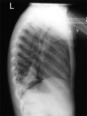
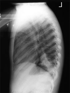
Lateral Chest X-ray
I thought there were universally accepted conventions for viewing lateral chest X-ray images.... but after doing a google images search, I'm not so sure.
Lateral Placement
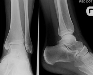
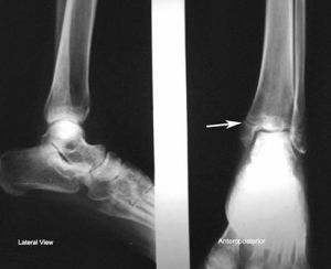
Two Views on a Single 'Film'- Orientation of the LateralSide-markers should be placed where they are least likely to obscure anatomy ( a do-no-harm philosophy). Some radiographers position side markers laterally with respect to the anatomy rather than medially. Once again, google images ( and K.C. Clarke) have lowered my confidence in this custom.
The first six AP ankle images from a google search followed the lateral side-marker placement custom and the following three didn't.
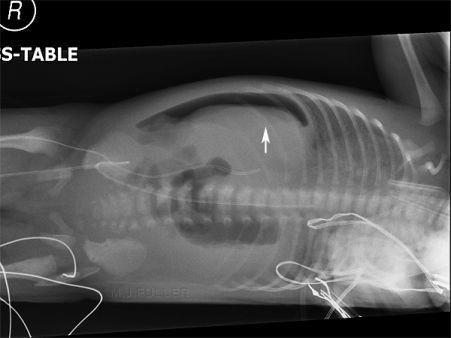
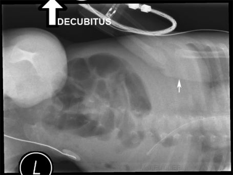
A further consideration with film/screen radiography is that the side-marker should be placed near the information/name window. This allows the radiologist to read all of the relevant information in the same location on the image.The Decubitus Side-marker Convention
This is the convention that is advocated in Merrill's and K.C. Clarke This is incorrect according to Merrill's and K.C. Clarke
Merrill's advice is as follows
"For decubitus positions of the chest and abdomen, the R or L marker should be placed on the side up (opposite the side laid on) and away from the anatomy of interest"
Radiograhic Positions and Radiologic Procedures, volume 1, tenth edition, p 25
One point that I believe is universally accepted is that, irrespective of side marking conventions used, a right side marker should never be placed on the patient's left side and vice versa.
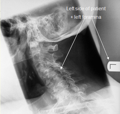
The Oblique Cervical Spine Labelling Convention
Two Views at Right Angles
Entry Site MarkerX-ray images present three dimensional anatomy in two dimensions. There should be a minimum of two projections at right angles for all imaged anatomy. Whilst there are a considerable number of exceptions, this principle is a very sound one and should always be considered, particularly in the Emergency Department where compromise and innovation in radiography are commonplace. Interestingly, conventional wrist radiographic technique results in a single view of the ulna- this is never mentioned in the radiography literature. The reason for this is that the radius rolls around a fixed ulna in pronation and supination.
At Least one Joint on all Long Bone X-ray ImagesIt is customary to mark the entry site (a lead shot dot is ideal) in cases of percutaneous foreign body where practical.
The Joint Above and the Joint BelowA long bone X-ray image that does not include at least one joint just looks wrong. More importantly, inclusion of at least one joint allows for the long bone to be identified and provides a reference point. Exceptions might include foreign body localisation where the foreign body provides the reference point and coned views of pathology where the pathology (and the routine views) provide the reference point.
Some consider that any long bone X-ray image should include two joints (joint above and joint below). This would require that, for example, all Colle's fractures should include the patient's elbow. I consider this rule to be inappropriate for a variety of reasons. Firstly, any anatomy should be imaged where there has been consideration of the cost vs the likely yield. To routinely image anatomy that is clinically normal is ludicrous (there are exceptions that should be assessed on their merits). In an effort to include the other normal joint, there will be distortion of the important anatomy associated with the oblique rays- a hand X-ray is not a wrist X-ray, is not a forearm X-ray, is not an elbow X-ray (irrespective of overlapping anatomy).
Customs and Practices in Digital Radiography
Will digital radiography spawn a whole new set of customs?
... back to the Applied Radiography home page
... back to the Wikiradiography home page
