Chest Anatomy
Jump to navigation
Jump to search
Introduction
Left Paraspinal Stripe and Descending Aorta
The Azygos Vein
... back to the wikiradiography home page
... back to the Applied Radiography home page
AcknowledgementThe chest X-ray is something of a holy grail for radiographers with an interest in image interpretation. A sound understanding of plain film chest anatomy provides a solid grounding for developing skills in chest X-ray image interpretation.
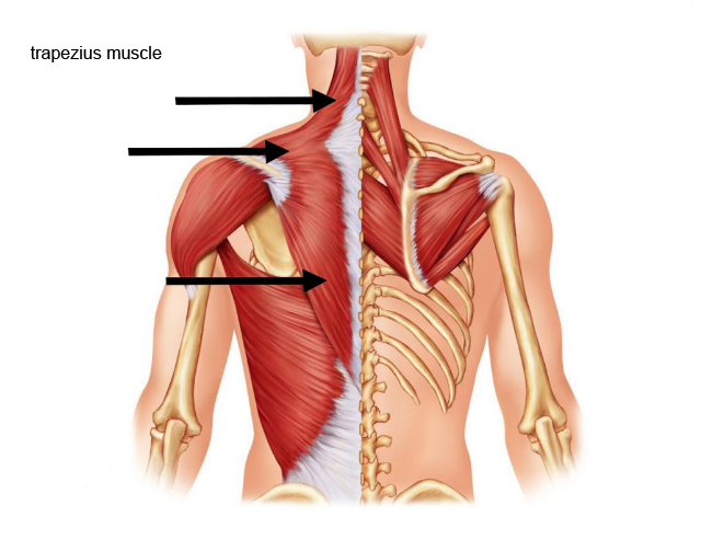
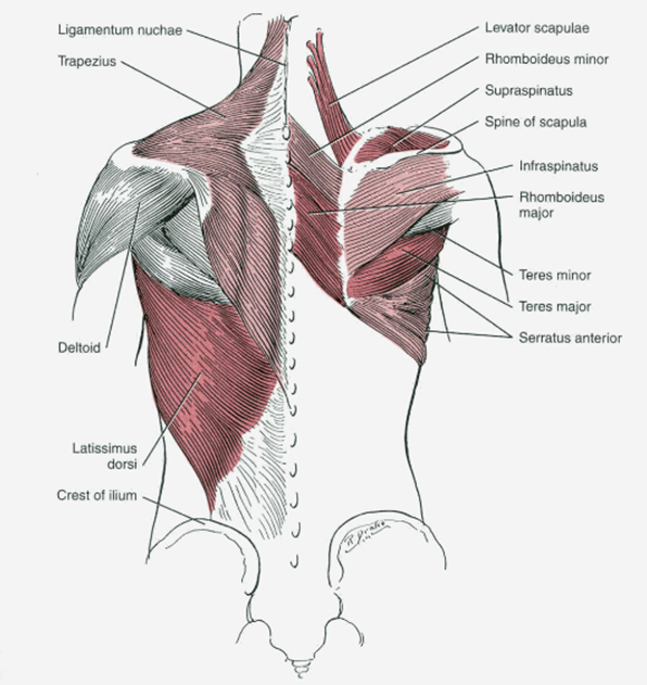
MusclesI have borrowed heavily from the work of Jerry M. Gibbs et al. This page is unlikely to do their original paper justice.
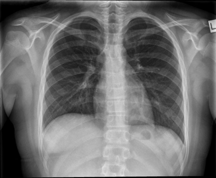
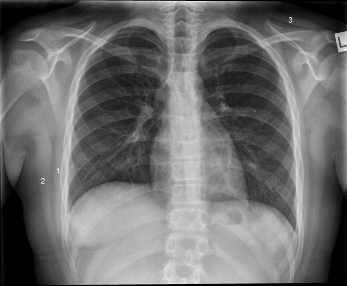
11 year old female with good demonstration of chest muscular anatomy
http://www.flashcardmachine.com/upper-body-muscles.html</a><a class="external" href="http://www.gainmuscleandloseweight.com/upper-back-workout/" rel="nofollow" target="_blank">
http://www.gainmuscleandloseweight.com/upper-back-workout/</a><a class="external" href="http://upload.wikimedia.org/wikipedia/commons/c/c9/Gray1215.png" rel="nofollow" target="_blank">
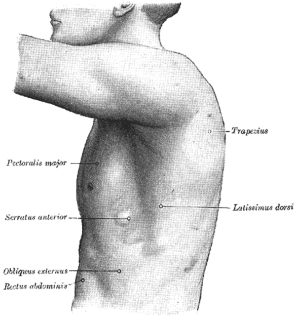
http://upload.wikimedia.org/wikipedia/commons/c/c9/Gray1215.png</a>1. Serratus Anterior
2. Latissimus dorsi
3. Trapezius
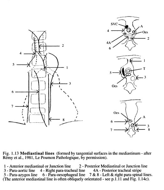
What are Chest X-ray Lines and Stripes?
There are various linear anatomical structures and borders within the mediastinum which have the potential to produce a linear density on a plain film chest X-ray image.
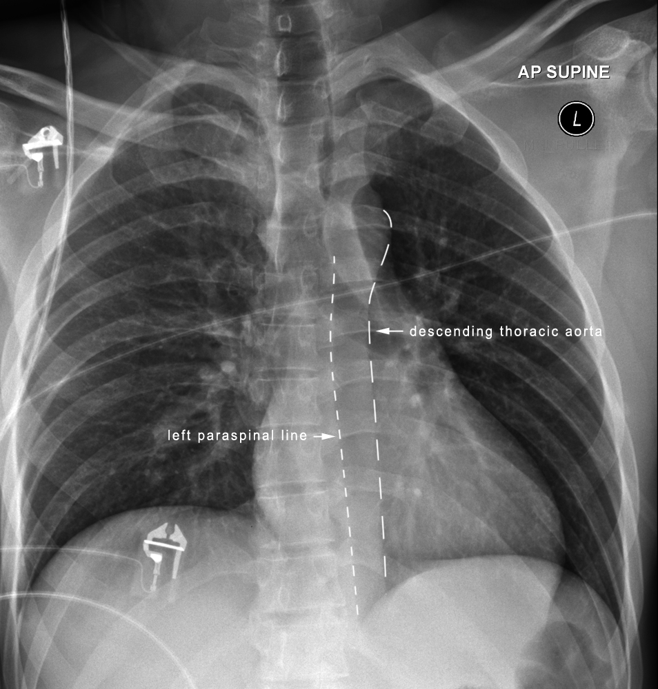
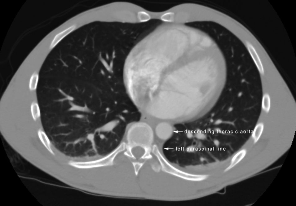
Left Paraspinal Stripe and Descending Aorta
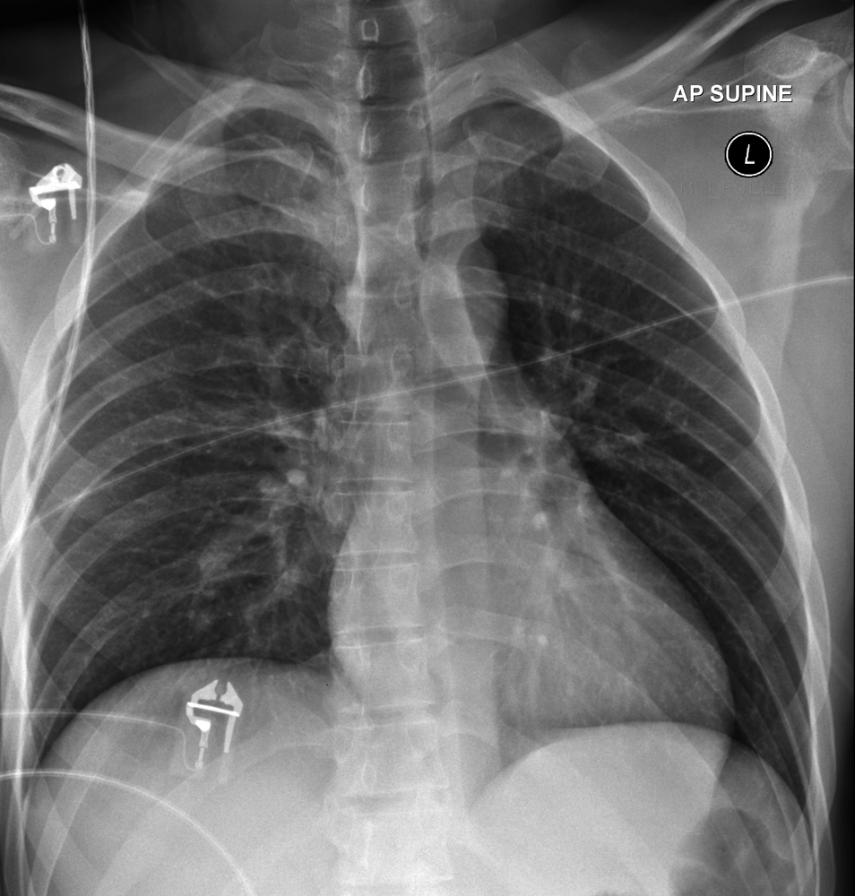
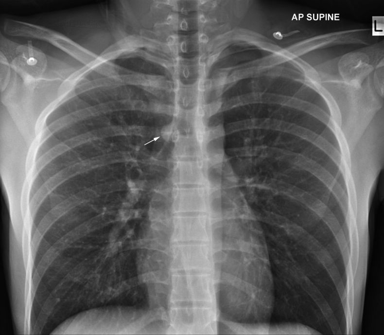
This AP supine chest X-ray is slightly LPO. The left paraspinal stripe and descending aorta are particularly well demonstrated. The left paraspinal line is formed by tangential contact of the left lung and pleura with the posterior mediastinal fat, left paraspinal muscles, and adjacent soft tissues.
<a class="external" href="http://radiographics.rsna.org/content/27/1/33.full.pdf" rel="nofollow" target="_blank">Jerry M. Gibbs, MD ● Chitra A. Chandrasekhar, MBBS ● Emma C.
Ferguson, MD ● Sandra A. A. Oldham, MD
Lines and Stripes:Where Did They Go?—From Conventional Radiography to CT
RadioGraphics 2007; 27:33–48</a>
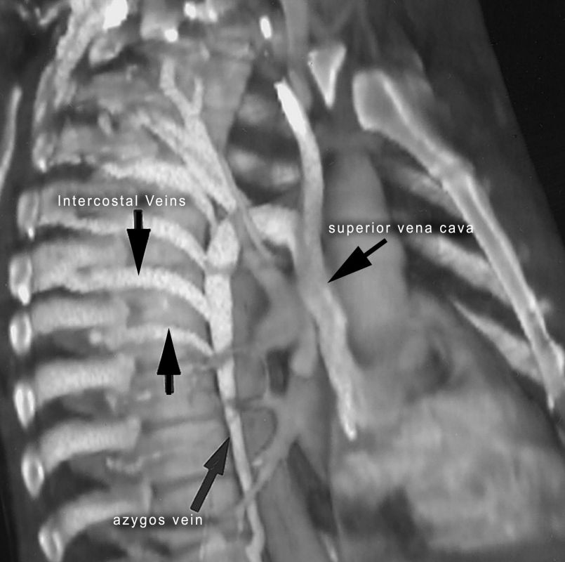
The Azygos Vein
<a class="external" href="http://radiographics.rsna.org/content/22/suppl_1/S45.full" rel="nofollow" target="_blank">
adapted from
Leo P. Lawler,Frank M. Corl, MS and Elliot K. Fishman, MD
Multi–Detector Row and Volume-rendered CT of the Normal and Accessory Flow Pathways of the Thoracic Systemic and Pulmonary Veins
RadioGraphics October 2002 vol. 22 no. suppl 1 S45-S60 </a>The azygos vein transports deoxygenated blood from the posterior walls of the thorax and abdomen into the superior vena cava . The vein is so named because it has no symmetrically equivalent vein on the left side of the body. It is formed by the union of the ascending lumbar veins with the right subcostal veins at the level of the 12th thoracic vertebra, ascending in the posterior mediastinum, and arching over the root of the right lung to join the superior vena cava. <a class="external" href="http://www.wikidoc.org/index.php/Azygos_vein" rel="nofollow" target="_blank">http://www.wikidoc.org/index.php/Azygos_vein</a> "The only part of the azygos system seen in the healthy chest is the arch of the azygos vein , which extends forward from the spine to its entry in the superior vena cava. Its end-on projection is seen on the frontal film as a round or oval shadow (arrowed) in the crotch between the takeoff of the RUL bronchus and the right tracheal border... It is invariably present in the recumbent position... however, it is frequently invisible on the erect film"
quoted from Chest Roentenology, Benjamin Felson, 1973, p232
... back to the wikiradiography home page
... back to the Applied Radiography home page
