--- under construction---
IntroductionUnexpected asymmetry of lung density can be perplexing for radiographer and clinician alike. This page considers the causes of lung density asymmetry and what can be done by the radiography to clarify the cause.
Common Causes of Asymmetrical Lung Density1. Pleural Space Air or Fluid
2. Technical
Grid cut-off
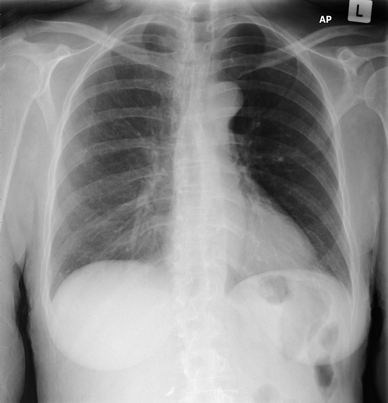 | This PA chest image demonstrates uneven lung density due to grid cut-off. There is a significant clue in that the left shoulder appears darker than the right shoulder. Importantly, this uneven demonstration of shoulder density matches the uneven lung density.This artifact can be interpreted as pathological. Insertion of an intercostal drain into the apparently hyperlucent hemithorax is not unheard of. |
Rotation
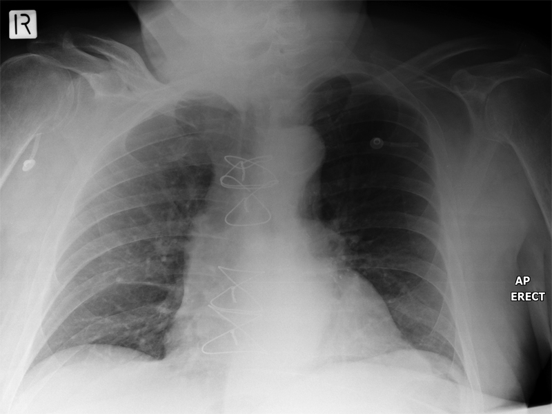 | This patient is in an AP sitting position. The CR cassette is positioned behind the patient's thorax. The patient has a left sided hemiplegia causing him to both lean and rotate. This is a relatively common scenario with bedside chest radiography. After several attempts to position the patient in an acceptable AP sitting position, the radiographers resorted to angling the beam and cassette to produce an AP chest position. This technique has resulted in an apparent uneven lung density. Note the shoulder positions and density. Note also the elongation of the right ribs.
The rotation of the chest, the asymmetrical shoulder positions, and the asymmetrical shoulder density are the clues for the radiologist that the apparent uneven lung density is artefactual rather than pathological. |
3. Uneven Aeration
ET tube malposition 1
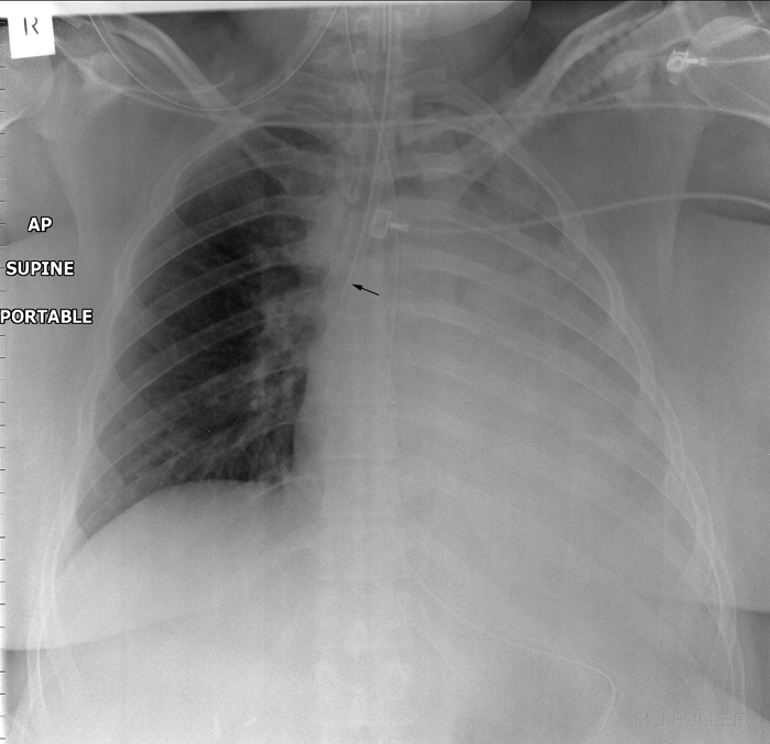 | This patient was referred for bedside chest radiography following intubation. There is almost complete loss of aeration of the left lung. The ET tube tip is positioned in the right main bronchus causing loss of aeration to the left lung. The left lung re-aerated when the ET tube was withdrawn. |
ET tube malposition 2
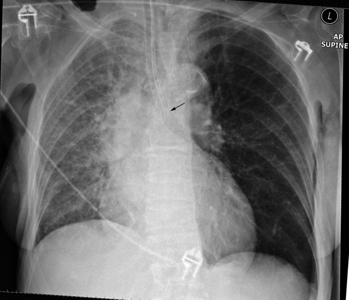 | The tip of the ET tube is in the left main bronchus. This si favouring aeration of the left lung at the expense of the right lung. |
Broncial Foreign Body
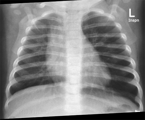 | This 7 month old boy presented to the Emergency Department after a choking episode. There was concern about an inhaled foreign body. Inspiration and expiration chest radiography was requested.
On inspiration the two lungs will tend to appear similar in terms of their degree of aeration. The reason for this is that the trachea and bronchii normally widen on inspiration allowing passage of air into the affected lung past the foreign body. |
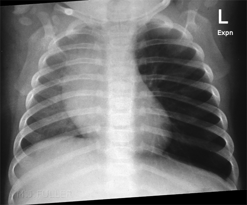 | On expiration the foreign body can obstruct the bronchii as the diameter of the bronchii decreases slightly. The greatest difference in lung aeration will therefore be seen on the expiration image as air is exhaled from the normal lung (right) but not from the affected lung(left). The pressure in the affected lung (left) remains relatively constant throughout respiration.
This child is likely to have a foreign body in the left main bronchus.
Note that in both inspiration and expiration images the diaphragms have not moved greatly. The heart and mediastinum are moving rather than the hemidiaphragms. |


