Carpal Tunnel Radiography
Jump to navigation
Jump to search
Introduction
Anatomy
Carpal Tunnel Syndrome
Radiography
What Went Wrong?
... back to the Applied Radiography home page
... back to the Wikiradiography home page
The carpal tunnel projection is rarely performed today. It is a projection that radiographers should be aware of and able to perform for those rarely encountered patients in whom it is indicated.
Anatomy
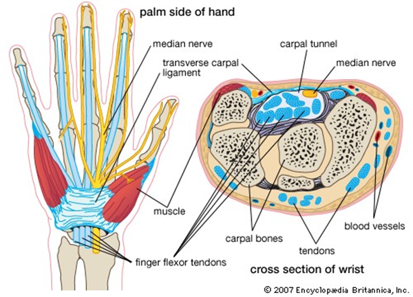
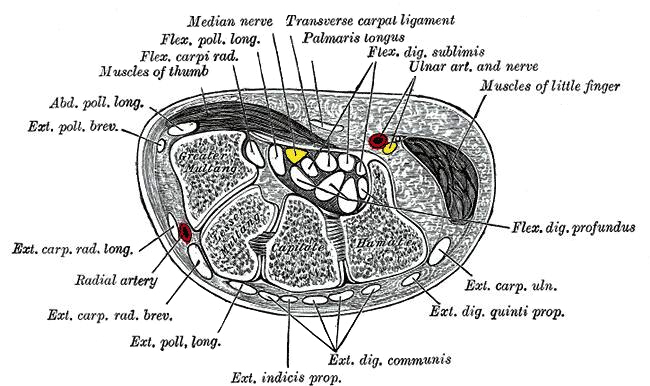
source: <a class="external" href="http://www.chiropractic-help.com/images/Carpal-tunnel1.jpg" rel="nofollow" target="_blank">http://www.chiropractic-help.com/images/Carpal-tunnel1.jpg</a>The carpal tunnel refers to the tunnel-like structure of the wrist in which 9 tendons and the median nerve pass through a narrow passage under a tight band of tissue known as the transverse carpal ligament. The transverse carpal ligament is also known as the flexor retinaculum or the anterior annular ligament. The carpal bones form a shallow concavity anteriorly, and the transverse carpal ligament completes the tunnel by its attachment medially to the pisiform and the hook of the hamate, and laterally to the tubercle of the scaphoid and the ridge of the trapezium
(Positioning in Radiography, 9th ed, K.C.Clarke, William Heinemann Medical Books LTD, London 1973 p24)
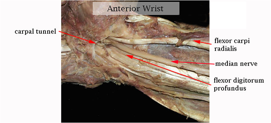
source: <a class="external" href="http://www.digherbs.com/image-files/carpal_tunnel_syndrome.jpg" rel="nofollow" target="_blank">http://www.digherbs.com/image-files/carpal_tunnel_syndrome.jpg</a>This image demonstrates the detailed carpal tunnel anatomy. Anatomical dissection demonstrating the relative sizes of the median nerve and the flexor carpi radialis.
Carpal Tunnel Syndrome
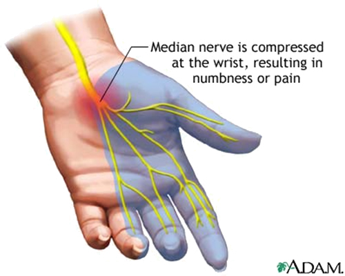
source: <a class="external" href="http://www.nlm.nih.gov/medlineplus/ency/images/ency/fullsize/1081.jpg" rel="nofollow" target="_blank">http://www.nlm.nih.gov/medlineplus/ency/images/ency/fullsize/1081.jpg</a>Carpal tunnel syndrome is a painful disorder of the hand caused by pressure on the median nerve which runs from the forearm to the hand passing through the carpal tunnel. Carpal tunnel syndrome symptoms include numbness, parasthesia (pins and needles), and pain (particularly at night). Anything that causes swelling inside the wrist can cause carpal tunnel syndrome, including repetitive hand movements, pregnancy and arthritis.<a class="external" href="http://www.steadyhealth.com/articles/Carpal_Tunnel_Syndrome_Prevention_a568.html#" rel="nofollow" target="_blank"></a> Carpal tunnel syndrome is one of the most common and widely known neuropathies in which the body's peripheral nerves are compressed or traumatized. Carpal tunnel syndrome is most often carpal tunnel syndrome is caused by repetitive motion, injury, or inflammatory types of arthritis. Treatment options include rest, splinting, cortisone injections and surgery.
Carpal tunnel radiography is generally not indicated for assessment of the carpal tunnel, and in particular has no role in the assessment of carpal tunnel syndrome (there may be rare exceptions). The volume, shape and architecture of the bony anatomy of the carpal tunnel is best evaluated with CT imaging. MRI and ultrasound imaging may be indicated for assessment of carpal tunnel pathology including carpal tunnel syndrome. Many surgeons consider the clinical signs of carpal tunnel syndrome sufficiently specific to negate the need for any imaging of the anatomy.
Radiography
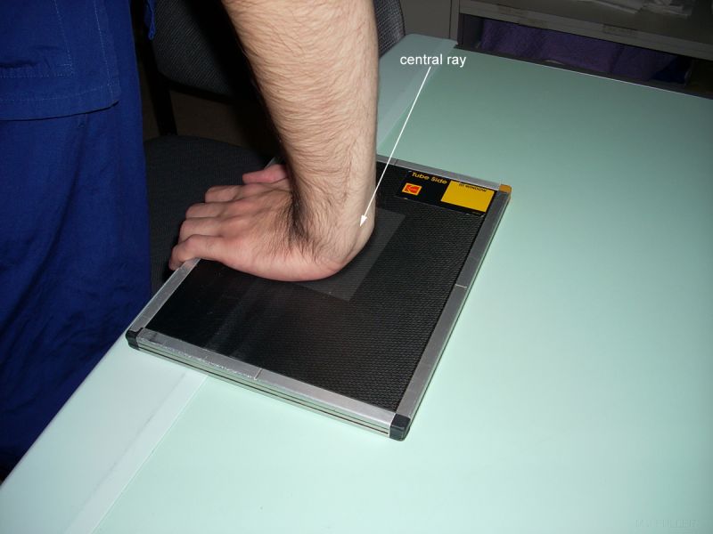
Technique 1
Technique 2
Technique 3
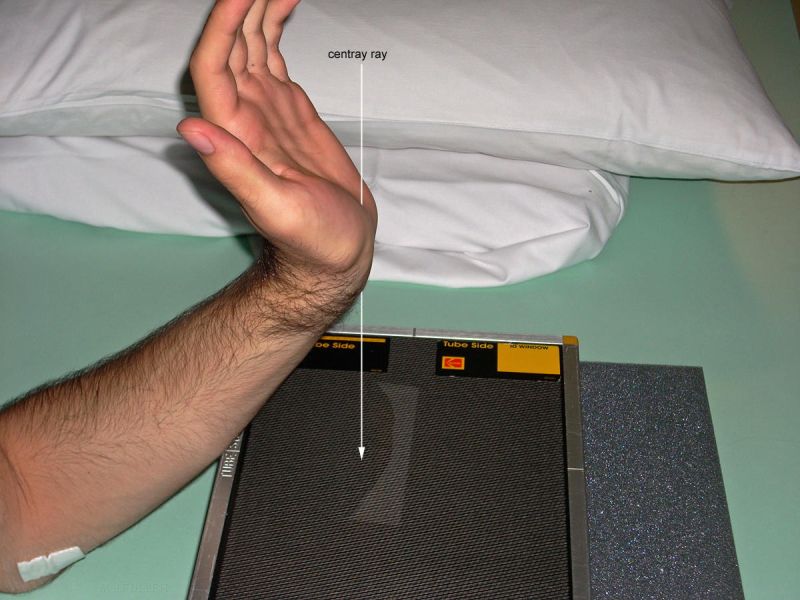
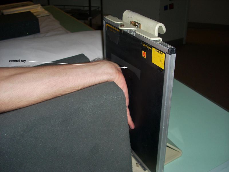
This is similar to the technique above. The additional forced dorsiflexion allows the forearm to be rested on the IR with tube angulation to suit the degree of hand dorsiflexion.
Technique 4
Technique 5
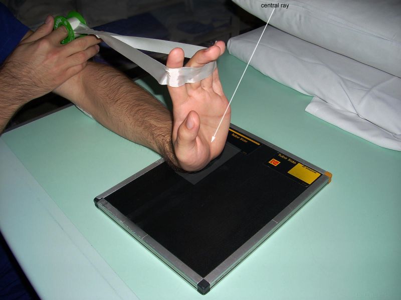
This is similar to the technique shown above. The patient's hand is in maximum dorsiflexion. The patient's forearm is supported with a DARRIN sponge.
Carpal Tunnel Radiography
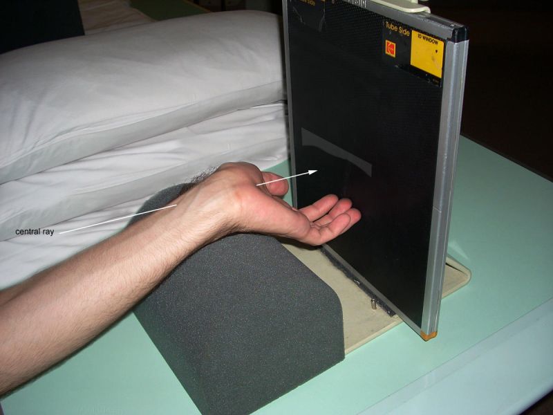
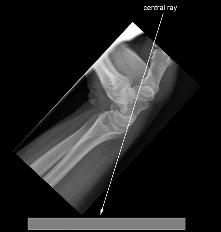
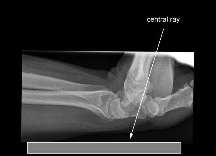
The carpal tunnel projection can be achieved using a variety of techniques. The X-ray beam can be vertical, angled or horizontal. The carpal tunnel projection image shown below was achieved with the patient's hand as shown left. If the patient cannot achieve sufficient hand dorsiflexion (wrist extension) , the examination will not be successful.
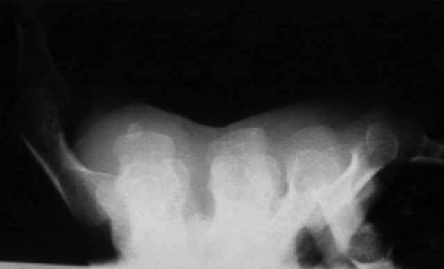
The Carpal Tunnel Projection Image
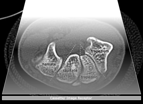
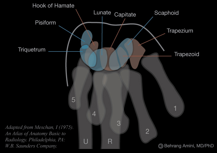
If the positioning is successful, the pisiform and hook of the hamate will be clearly demonstrated. The carpal bones will also be displayed as a tunnel.
The Radiographic Anatomy of the Carpal Tunnel Projection
<a class="external" href="http://radiopaedia.org/uploads/radio/0013/0253/carpal-tunnel-view.jpg" rel="nofollow" target="_blank">
http://radiopaedia.org/uploads/radio/0013/0253/carpal-tunnel-view.jpg</a>
What Went Wrong?
Case 1
Case 2
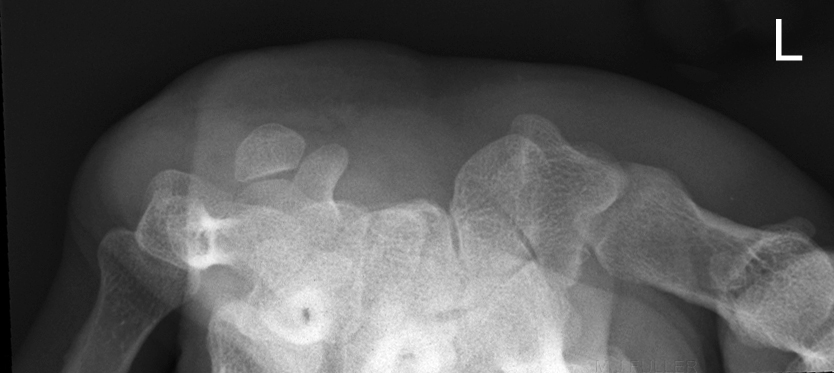
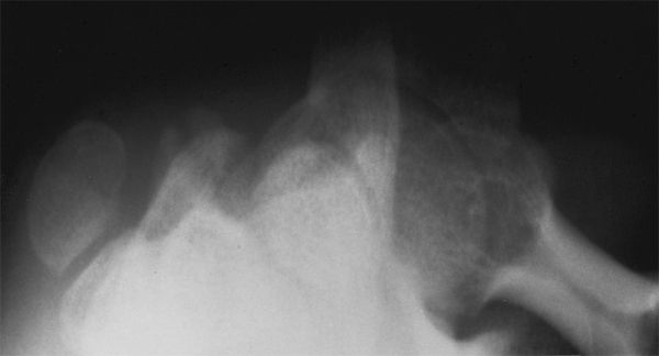
adapted from <a class="external" href="http://radiology.rsna.org/content/219/1/11/F39.large.jpg" rel="nofollow" target="_blank">http://radiology.rsna.org/content/219/1/11/F39.large.jpg</a>I suspect that this patient has excessive dorsiflexion of the hand. There may also be a rotation error. Fracture of the hook of the hamate noted.
... back to the Applied Radiography home page
... back to the Wikiradiography home page
