CT Case 4 - Le Fort Fractures
Jump to navigation
Jump to search
Introduction
A CT of the head and facial bones was performed on a 43 year old female that presented with severe facial trauma following a fall from 30 metres.
Results
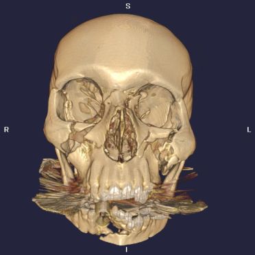
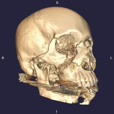
There are extensive fractures of the facial bones that affect all bones of the face except the hard palate. This appearance reflects complex Le Fort I, II, and III fractures. See Image A and Image B below.
Discussion
Facial fractures can be broadly classified using the Le Fort system which was devised by Paris surgeon Renee Le Fort in 1901.
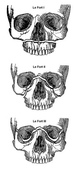
Le Fort I - fracture is transverse and separates the tooth-bearing part of the maxilla from the rest of the maxilla
Le Fort II - fracture extends through the medial and lateral walls of the maxillary sinuses and through the nasal bones resulting in a pyramid shaped fracture fragment
Le Fort III - the facial bones are completely separated (craniofacial dysjunction) from the skull with fractures through the medial and lateral walls of the orbits and through the zygomatic arches
Multiple fractures can be present and the different fracture types may co-exist. Le Fort I, II, and III fractures are illustrated by Image C.
References
Moore, K (1992) ‘Clinically oriented anatomy’, 3rd edn, Williams & Wilkins, Baltimore.
Tennessee Craniofacial Center, Chapter 8: Maxillary Fractures, <a class="external" href="http://www.craniofacialcenter.com/book/trauma/trauma_3.htm" rel="nofollow" target="_blank">http://www.craniofacialcenter.com/book/trauma/trauma_3.htm</a>, accessed 20 March 2007.
Weir, J & Murray, A (1998) ‘Mosby’s atlas and text of clinical imaging’, Mosby-Wolfe, London.
Introduction
A CT of the head and facial bones was performed on a 43 year old female that presented with severe facial trauma following a fall from 30 metres.
Results
There are extensive fractures of the facial bones that affect all bones of the face except the hard palate. This appearance reflects complex Le Fort I, II, and III fractures. See Image A and Image B below.

Image B 3D reconstruction of oblique skull demonstrating Le Fort facial fractures.

Image A 3D reconstruction of AP skull demonstrating Le Fort facial fractures.
Discussion
Facial fractures can be broadly classified using the Le Fort system which was devised by Paris surgeon Renee Le Fort in 1901.
Le Fort I - fracture is transverse and separates the tooth-bearing part of the maxilla from the rest of the maxilla
Le Fort II - fracture extends through the medial and lateral walls of the maxillary sinuses and through the nasal bones resulting in a pyramid shaped fracture fragment
Le Fort III - the facial bones are completely separated (craniofacial dysjunction) from the skull with fractures through the medial and lateral walls of the orbits and through the zygomatic arches
Multiple fractures can be present and the different fracture types may co-exist. Le Fort I, II, and III fractures are illustrated by Image C.

Image C Le Fort I (top), Le Fort II (middle), and Le Fort III (bottom) fractures (Tennessee Craniofacial Centre)
References
Moore, K (1992) ‘Clinically oriented anatomy’, 3rd edn, Williams & Wilkins, Baltimore.
Tennessee Craniofacial Center, Chapter 8: Maxillary Fractures, <a class="external" href="http://www.craniofacialcenter.com/book/trauma/trauma_3.htm" rel="nofollow" target="_blank">http://www.craniofacialcenter.com/book/trauma/trauma_3.htm</a>, accessed 20 March 2007.
Weir, J & Murray, A (1998) ‘Mosby’s atlas and text of clinical imaging’, Mosby-Wolfe, London.