IntroductionComputed radiography (CR) provided the world of radiography with an intermediate step between film/screen radiography and direct digital (DR) imaging systems. Many imaging departments will have both technologies... even in the same room. This page considers anecdotal evidence regarding the image quality differences between CR and DR imaging- is the difference clinically significant?
Case 1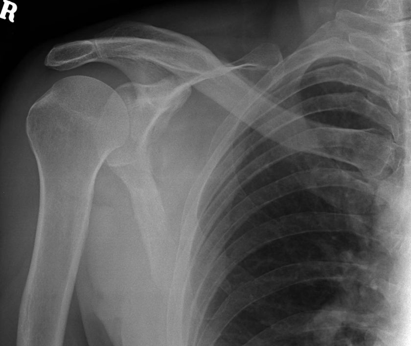 | This 61 year old male presented to the Emergency Department after falling backwards while dancing. He was examined and found to have a painful right scapula. He was referred for right shoulder radiography.
The radiographer noted the high level of pain and discomfort that the patient was demonstrating and therefore chose to utilise a CR non-grid bedside approach to the shoulder radiography. This was a reasonable judgement and reflects an approach that many (if not most) radiographers would have employed in the same circumstances.
The AP shoulder image demonstrates no obvious displaced fracture. |
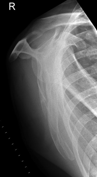 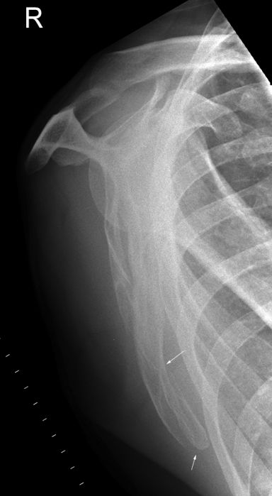 | The AP supine rolled lateral scapula projection image demonstrates a fracture involving the inferior angle of the scapula.
Given that the clinical question had been answered and the radiographer had demonstrated the fracture, is any further imaging justified?
It is not unusual for experienced radiographers to be able to identify fractures which are not detected by the doctors in the Emergency Department. In some cases, the fractures are also missed by the reporting radiologist. You could argue that this is not the radiographer's problem- they have done their job. It is of course little succour to the patient that has been discharged and advised that they have no fracture and can expect to feel better in a few days with minimal analgesia! |
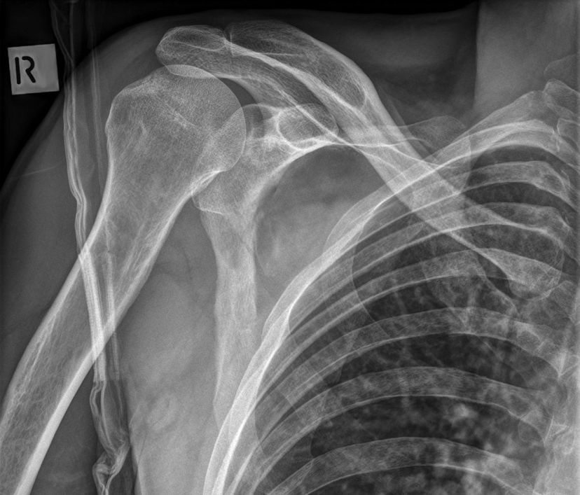 | Further imaging of the patient's right shoulder was undertaken utilising a DR system. |
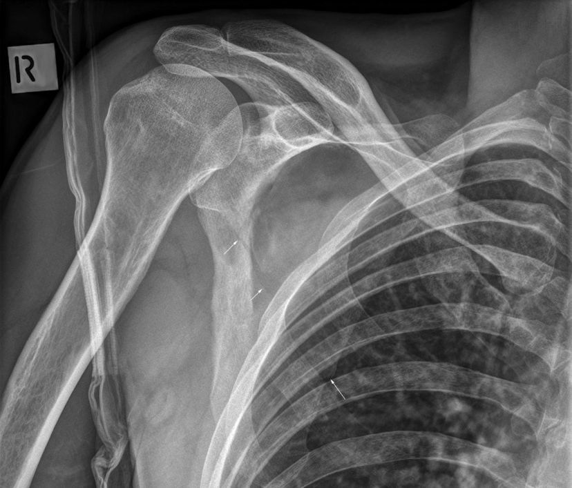 | The patient was unable to rotate further. A long exposure time (0.8 seconds was the maximum exposure time available with this DR system) was employed and the patient was breathing during the exposure- this did achieve some bluring (movement unsharpness) of the lung markings resulting in improved demonstration of the scapula. The fractured scapula was demonstrated clearly with additional evidence of the fracture extending up towards the glenoid (arrowed). In retrospect, this was also demonstrated on the original CR AP shoulder image.
mattress artifact noted |
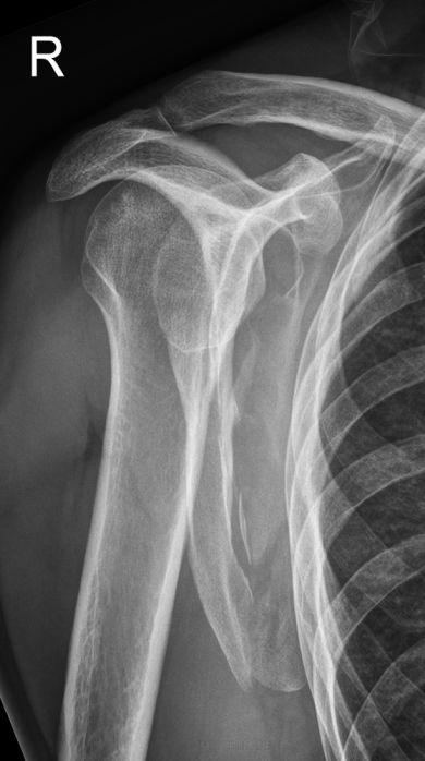 | This is a deliberate under-rotated lateral scapular position- this oblique shoulder position can be very useful in demonstrating scapular fractures. This is a DR image. |
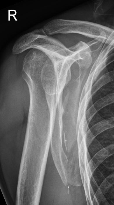 | The DR oblique shoulder image demonstrates the fracture of the inferior aspect of the scapula clearly. |
Comment
Where there are both bucky DR and cassette CR systems available, the radiographer must make a balanced judgement as to which system is most appropriate given all of the circumstances of the case. The DR system is more likely to produce images which demonstrate all fractures clearly. Beware satisfaction syndrome.
Case 2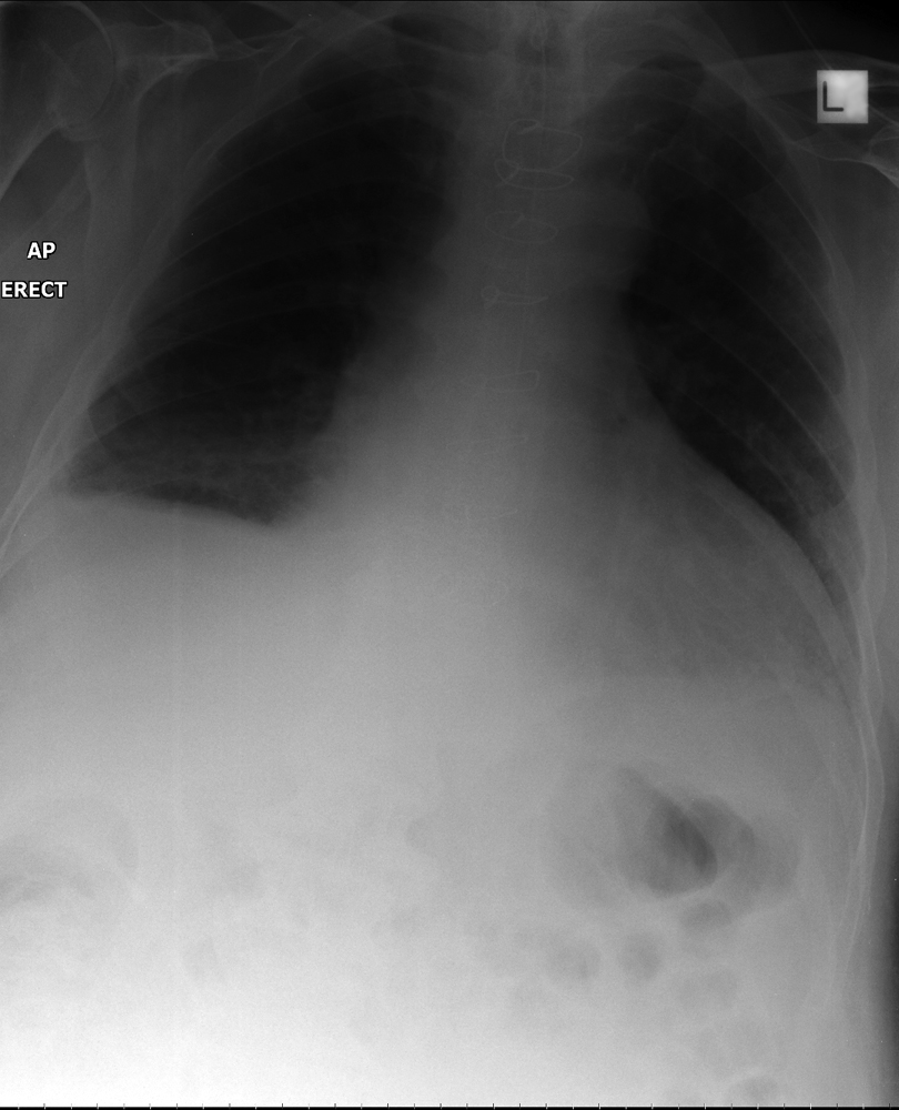 | This 82 year old man was found to have a non-draining NGT and was referred for chest radiography. The radiographer considered the patient to be too unwell to be subjected to PA erect chest radiography and undertook AP sitting CR technique. The NGT could be visualised proximally, but the tip position was unclear. In order to demonstrate the tip position the options included DR technique (erect or supine) or using the same CR technique with a stationery grid. THe DR option was chosen. |
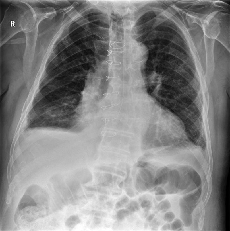 | The erect PA DR image demonstrates the NGT tip position to be in the distal third of the oesophagus.
- sternal sutures noted.
- bilateral subpulmonic effusions noted, larger on the right |
... back to the
wikiradiography home page
... back to the
Applied Radiography home page


