Bone Pathology
Jump to navigation
Jump to search
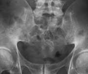
In younger patients Ankylosing Spondylitis may affect the spine and pelvis. Fusion of the scaroiliac joints precedes spinal involvement.
Yaweta Ndovi
Osteopetrosis - Submitted by Kelly Spence
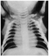
This is an image of osteopetrosis which is caused by a lack of bone Calcium reabsorbtion.
----------------------------------------------------------------------------------------------------------------------------------------------------------------------------------
This is a genetics study patient who has a rare condition called Osteopetrosis. Shes is
being studied because her son has also been diagnosed with the same condition.
MOTHERs Lateral: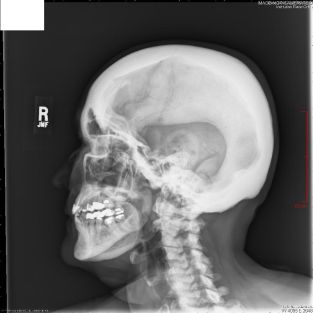 MOTHERS AP:
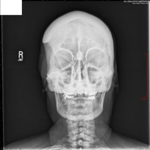
SON's Lateral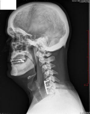 SON's AP
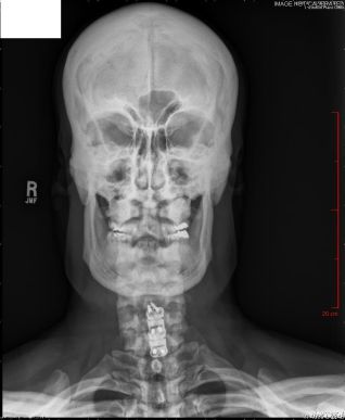

In younger patients Ankylosing Spondylitis may affect the spine and pelvis. Fusion of the scaroiliac joints precedes spinal involvement.
Yaweta Ndovi
| |  Ankylosing Spondylitis. |
 Eosinophilic Granuloma (EG) EG is a lesion with many histiocystes that may contain Langerhans granules & pockets of necrosis. Usually is a solitary bone lesion. The one in this x-ray is in the distal humeral shaft of a 14 yr.old. Eosinophilic Granuloma (EG) EG is a lesion with many histiocystes that may contain Langerhans granules & pockets of necrosis. Usually is a solitary bone lesion. The one in this x-ray is in the distal humeral shaft of a 14 yr.old. |  Rheumatoid Arthritis is a chronic systemic disease causing inflammation of small joints of hands and feet. |
 Paget's disease is a chronic metabolic disease of the skeleton. It's commonly seen during middle life and it affects men twice as often as women. In this radiograph, tibia is in the reparative stage which is characterized by enlarged bone, cortical thickening and coarse trabeculae. Paget's disease is a chronic metabolic disease of the skeleton. It's commonly seen during middle life and it affects men twice as often as women. In this radiograph, tibia is in the reparative stage which is characterized by enlarged bone, cortical thickening and coarse trabeculae. |  Gout An acute inflammatory disease characterized by the build up of uric acid. This causes crystals to form on the articular cartilage of joints, tendons, and surrounding tissues. The patient usually suffers from excruciating, sudden, unexpected, burning pain, swelling, redness, warmness and stiffness in the joint. Gout An acute inflammatory disease characterized by the build up of uric acid. This causes crystals to form on the articular cartilage of joints, tendons, and surrounding tissues. The patient usually suffers from excruciating, sudden, unexpected, burning pain, swelling, redness, warmness and stiffness in the joint.Swoodlee |
 Chondrosarcoma is a type of malignant bone cancer that develops in the cartilage cells. It primarily affects the femur, knee, arm, pelvis and spine. It is the second most common type of bone cancer and is usually found in individuals between the ages of 50 and 70. Its etiology is unknown. S.Jones |  Ewing's sarcoma occurs most frequently in male teenagers. It is found most commonly in the pelvis and proximal long tubular bones. Tplank |
| A Giant cell tumor of the distal femur - an aggressive benign tumor | Osteoid osteoma of the coracoid process of the scapula (arrow). posted by Gulilat Yimenu  |


Osteopetrosis - Submitted by Kelly Spence

This is an image of osteopetrosis which is caused by a lack of bone Calcium reabsorbtion.
----------------------------------------------------------------------------------------------------------------------------------------------------------------------------------
This is a genetics study patient who has a rare condition called Osteopetrosis. Shes is
being studied because her son has also been diagnosed with the same condition.
MOTHERs Lateral:
 MOTHERS AP:
SON's Lateral
 SON's AP