Acute Abomen1
This patient presented to the Emergency Department with acute abdominal pain. The patient's history included appendectomy,
Imaging
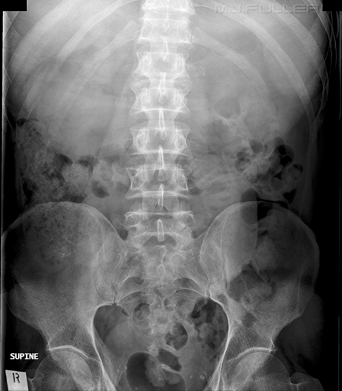
The patient was referred for abdominal imaging and the radiographer performed a supine abdominal X-ray examination. This image was reviewed to asses whether further abdominal imaging was required.
What pathology, if any, was demonstrated and are there reasonable grounds for performing additional views of the abdomen?
Findings
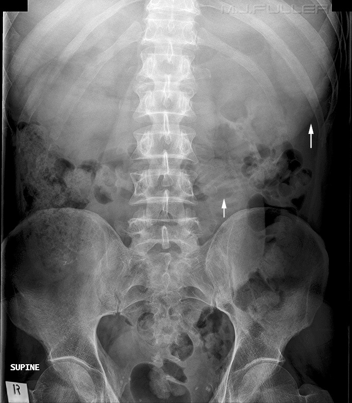
The radiographer noted that there appeared to be significant abnormal density extending across the upper abdomen.
The arrowed structure (upper arrow) was thought to represent the greater curve of the stomach. The appearances suggested a distended fluid-filled stomach positioned across the upper abdomen.
In addition, a dilated fluid filled loop of jejunum was noted with evidence of stretch sign. There was otherwise a paucity of small bowel gas.
The finding suggested to the radiographer that the patient could have a proximal small bowel obstruction (SBO).
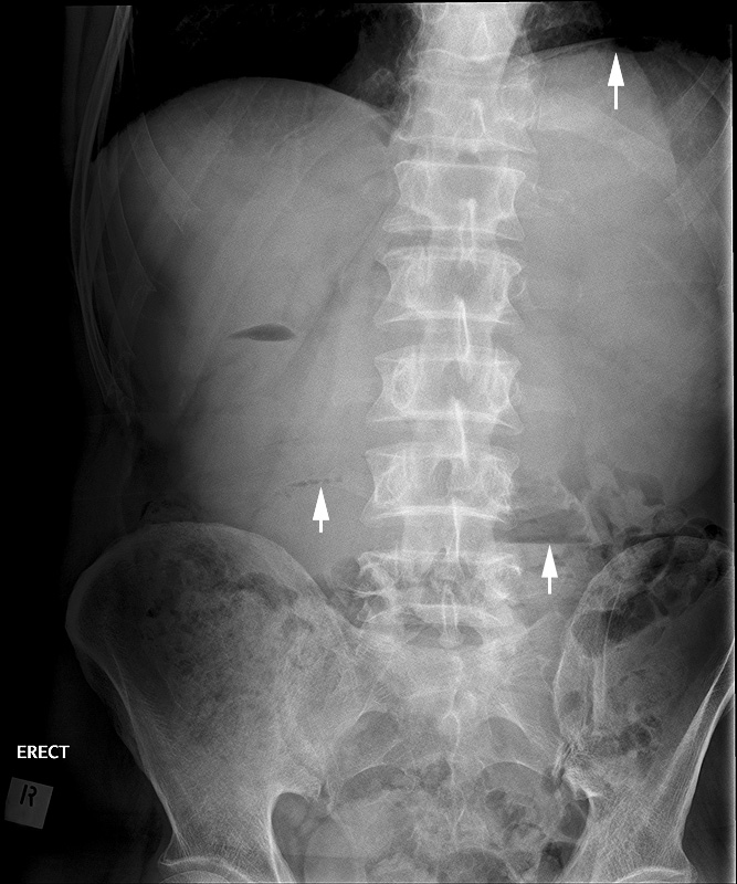
The radiographer considered that an erect abdominal image might provide additional useful information to help confirm the presence of a SBO.
The erect abdominal film also suggested a distended fluid-filled stomach. The top arrow points to a crescent of air in a fluid-filled gastric fundus.
There is a small string-of-pearls sign suggesting SBO (middle arrow).
There is also an air-fluid level in what appears to be a dilated loop of jejunum.
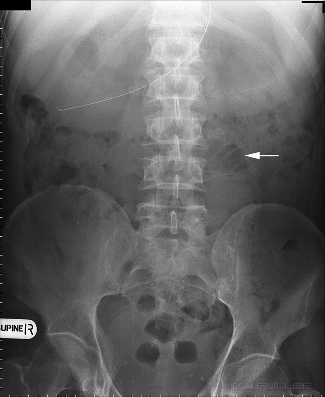
The patient was treated with a naso-gastric tube and repeat abdominal plain film imaging was performed. The dilated stomach now contains air and fluid (not arrowed) and the stretch sign is again demonstrated.
Discussion
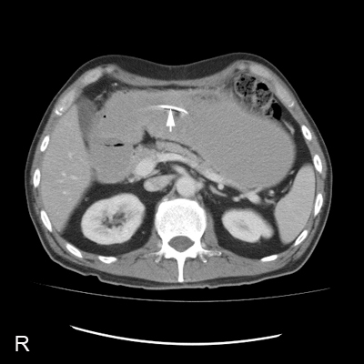
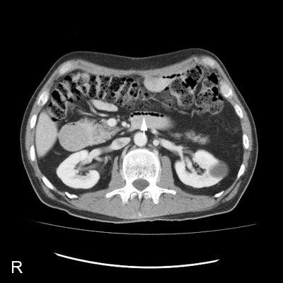
The patient was referred for a CT of the abdomen. This axial slice demonstrates the dilated fluid-filled stomach with NGT insitu (arrowed). This image demonstrates a few fluid-filed dilated loops of jejunum and a string-of -pearls sign (arrowed).
This case demonstrates the value of radiographers developing abdominal plain film pattern recognition skills. The radiographer's skills in considering the clinical presentation and identifying the stretch sign provided a strong indication for an erect abdominal plain film. It could be argued that a diagnosis of SBO could be made by a Radiologist on the supine image alone. This is of little utility to the patient or the referring doctor in the middle of the night when the radiologist is home asleep. The patient received a timely insertion of a NGT because of the radiographers successful demonstration of a SBO.
The abdominal plain film can be perceived by radiographers as a mundane aspect of radiography. I would argue that this need not be the case. A "clinical approach" rather than a "photographic approach" to radiography is more satisfying and effective.
...back to the Applied Radiography home page
