Acromioclavicular Joint Radiography
Introduction
Acromioclavicular joint (AC) injuries are commonly seen in Emergency Departments. This page considers all aspects of AC joint Radiography
Clinical Appearances
<embed allowfullscreen="true" height="350" src="http://widget.wetpaintserv.us/wiki/wikiradiography/widget/youtubevideo/fb4aad06d83f99b2a38c12b02a6755c355c90337" type="application/x-shockwave-flash" width="425" wmode="transparent"/> <embed allowfullscreen="true" height="350" src="http://widget.wetpaintserv.us/wiki/wikiradiography/widget/youtubevideo/520d73fddb7797c5adb2f91d938857f0b894aee2" type="application/x-shockwave-flash" width="425" wmode="transparent"/> <embed allowfullscreen="true" height="350" src="http://widget.wetpaintserv.us/wiki/wikiradiography/widget/youtubevideo/5d9b49d27318443b396f3d59081532935e5d5aa6" type="application/x-shockwave-flash" width="425" wmode="transparent"/> <embed allowfullscreen="true" height="350" src="http://widget.wetpaintserv.us/wiki/wikiradiography/widget/youtubevideo/96003e504065e4ea34dc8921d14dc4519ce612ae" type="application/x-shockwave-flash" width="425" wmode="transparent"/>
source: <a class="external" href="http://1.bp.blogspot.com/_teiZk-aADB4/SRI9s4kdufI/AAAAAAAAANQ/fvMLnf9wd_g/s1600-h/IMG_3382.jpg" rel="nofollow" target="_blank">http://1.bp.blogspot.com/_teiZk-aADB4/SRI9s4kdufI/AAAAAAAAANQ/fvMLnf9wd_g/s1600-h/IMG_3382.jpg</a>An AC joint subluxation has a characteristic appearance of a 'bump' in the contour of the shoulder. Compare the appearances of a subluxed AC joint with an anterior dislocation of the glenohumeral joint.


Grading an AC Joint Injury
RadiographyMost AC joint separations can be graded I, II or III, (mild, moderate or severe). There are actually six grades of severity of AC joint separations but grades four through six generally require extensive trauma as from a vehicular accident. A grade I separation is the most common and is characterized by a stretching or mild tearing of the acromioclavicular ligament. Other ligaments are unaffected and the collarbone does not lift as does with a more significant injury. With a grade II injury, the acromioclavicular ligament is usually completely torn and the coracoclavicular ligaments may be stretched. The collarbone will lift somewhat although this may not be evident on examination due to swelling around the A-C joint which will obscure the elevated collarbone. A grade III injury is more severe with complete tearing of the acromioclavicular ligament as well as significant tearing of the coracoclavicular ligaments. This lack of support between the shoulder blade and the collarbone causes the shoulder complex to drop and the collarbone to lift.<a class="external" href="http://www.theacc.com/genrel/120905aad.html" rel="nofollow" target="_blank"> Stephen Bushee, ATC, Assistant Athletic Director - Sports Medicine, Head Athletic Trainer, Boston Colleg, Acromioclavicular Separation in Ice Hockey, Typical injury...different mechanism!</a>
Normal AC Joint
<a class="external" href="http://www.leadingmd.com/patientEd/shoulder5/overview.asp" rel="nofollow" target="_blank">http://www.leadingmd.com/patientEd/shoulder5/overview.asp</a>Normal AC Joint An AC joint dislocation can involve injury to the coracoclavicular ligament as well as the acromioclavicular ligament. It is important radiographically to demonstrate the distance between the acromion and the clavicle.
<a class="external" href="http://www.theacc.com/genrel/120905aad.html" rel="nofollow" target="_blank"> Stephen Bushee, ATC, Assistant Athletic Director - Sports Medicine, Head Athletic Trainer, Boston Colleg, Acromioclavicular Separation in Ice Hockey, Typical injury...different mechanism!</a>Grade I AC Joint Separation
<a class="external" href="http://www.leadingmd.com/patientEd/shoulder5/overview.asp" rel="nofollow" target="_blank">http://www.leadingmd.com/patientEd/shoulder5/overview.asp</a>Grade I AC Joint Separation A grade I separation is the most common and is characterized by a stretching or mild tearing of the acromioclavicular ligament. Other ligaments are unaffected and the collarbone does not lift as does with a more significant injury.
<a class="external" href="http://www.theacc.com/genrel/120905aad.html" rel="nofollow" target="_blank"> Stephen Bushee, ATC, Assistant Athletic Director - Sports Medicine, Head Athletic Trainer, Boston Colleg, Acromioclavicular Separation in Ice Hockey, Typical injury...different mechanism!</a>Grade II AC Joint Separation
<a class="external" href="http://www.leadingmd.com/patientEd/shoulder5/overview.asp" rel="nofollow" target="_blank">http://www.leadingmd.com/patientEd/shoulder5/overview.asp</a>Grade II AC Joint Separation With a grade II injury, the acromioclavicular ligament is usually completely torn and the coracoclavicular ligaments may be stretched. The collarbone will lift somewhat although this may not be evident on examination due to swelling around the A-C joint which will obscure the elevated collarbone.
<a class="external" href="http://www.theacc.com/genrel/120905aad.html" rel="nofollow" target="_blank"> Stephen Bushee, ATC, Assistant Athletic Director - Sports Medicine, Head Athletic Trainer, Boston Colleg, Acromioclavicular Separation in Ice Hockey, Typical injury...different mechanism!</a>Grade III AC Joint Separation
<a class="external" href="http://www.leadingmd.com/patientEd/shoulder5/overview.asp" rel="nofollow" target="_blank">http://www.leadingmd.com/patientEd/shoulder5/overview.asp</a>Grade III AC Joint Separation A grade III injury is more severe with complete tearing of the acromioclavicular ligament as well as significant tearing of the coracoclavicular ligaments. This lack of support between the shoulder blade and the collarbone causes the shoulder complex to drop and the collarbone to lift.
<a class="external" href="http://www.theacc.com/genrel/120905aad.html" rel="nofollow" target="_blank">Stephen Bushee, ATC, Assistant Athletic Director - Sports Medicine, Head Athletic Trainer, Boston Colleg, Acromioclavicular Separation in Ice Hockey, Typical injury...different mechanism!</a>
"The Coracoclavicular ligaments–“Suspensory ligaments of the upper extremity”–Two components:•Trapezoid•Conoid–Stronger than AC ligaments–Provide vertical stability to AC joint"
Source: Injuries of the Clavicle, Acromioclavicular Joint and Sternoclavicular Joint
Andrew H. Schmidt, M.D., T.J. McElroy, January 2007
<a class="external" href="http://www.scribd.com/doc/21709781/U01-Clavicle-AC-SC-Joints" rel="nofollow" target="_blank">U01 clavicle AC SC Joints 1.ppt</a>


The AP view of the AC joints can be performed with or without cephalic tube angulation. The advantage of performing the AP view with cephalic tube angulation is that the AC joint is projected clear of the more proximal aspect of the acromion.
Both AC joints should always be imaged- this is one of those rare occasions where a comparison view is routinely performed. Whilst there may be exceptions to this rule (such as follow-up imaging of a known pathology), they should be rare events.
Binocular Cone for Acromioclavicular joints
The AC joints can be imaged with a binocular cone. This one was made by the author using 1mm lead glued between two sheets of laminex. The hole size and spacings can be determined using a mockup made of cardboard (or similar). The cone is attached to a piece of polycarbonate that slides into the rails on the LBD. The binocular cone is a special purpose cone for acromio-clavicular joint imaging. In one exposure you can image both AC joints without primary beam exposure to the patient's thyroid. By varying the FFD/SID the circles become larger and further apart.
The binocular cone will produce an image of the AC joints that looks like this.


Separate Exposure Method
The AC joints can be exposed separately as shown left. This method minimises the radiation dose to the patient's thyroid but can sometimes result in non-comparable projections- imaging of both joints in a single exposure tends to produce more comparable demonstration of the AC joints.
These AJ joints were imaged using the 2 exposure method. These are radiographs (as opposed to digital radiography images) demonstrating the difficulty in achieving a uniform exposure across the image.


Weight-bearing Views
[view AC joint&source=bl&ots=CByK2XpE7G&sig=OhEvbLdTa6-ajD954fGfDDiNZGI&hl=en&ei=Ww0NS5SPOoPxkAXwzICFBA&sa=X&oi=book_result&ct=result&resnum=4&ved=0CBcQ6AEwAw#v=onepage&q=zanca view AC joint&f=false|Source : Examination and Diagnosis of Musculoskeletal Disorders. ]
[view AC joint&source=bl&ots=CByK2XpE7G&sig=OhEvbLdTa6-ajD954fGfDDiNZGI&hl=en&ei=Ww0NS5SPOoPxkAXwzICFBA&sa=X&oi=book_result&ct=result&resnum=4&ved=0CBcQ6AEwAw#v=onepage&q=zanca view AC joint&f=false|Clinical Examination/Imaging Modalities.John R. Denton, MD ]Stress views of the AC joints may demonstrate instability and differentiate grade III AC separations from partial Grade I-II injuries. They are performed by having patient hold weights (e.g. 5 litre plastic bottles filled with water or sand). Both sides are imaged for comparison. Weight-bearing AC joint radiography is rarely undertaken today because most AC joint injuries are treated the same.
•Rationale: will demonstrate instability and differentiate grade III AC separations from partial Grade I-II injuries•Performed by having patient hold weight with injured arm•Rarely used today, since most AC joint injuries treated the same

The Zanca View


The Special Stress Views
These are a series of views to assess stability of an AC joint separation. The patient's right arm is in maximum adduction in this view.
The patient's arm is behind his/her back in this view This is possibly one of the few occasions where a weightbearing view of the AC joint can be justifiably performed without the contralateral comparison view. The reasoning is that the comparison is with the neutral view of the same side rather than the other side.


The Hidden AC Joint Injury
There have been numerous occasions where I have seen patients with obvious AC joint subluxations clinically, with little or no evidence of AC joint subluxation radiographically.
This patient could be seen to have a high grade AC joint subluxation (at least grade III) with no evidence of this injury radiographically. This is a supine AP shoulder. This is an unsuitable technique for assessment of AC joint injury.
The radiographer performed erect AC joint views as shown below.The injury to the left AC joint (grade III subluxation) was obvious when the patient was imaged in the erect position. The erect imaging could be considered to be a form of "weight-bearing" AC joints in that the weight of the patients arm will tend to reveal a subluxed AC joint.


What Went Wrong?
This looks like a subacromial view- the AC joint is superimposed over the acromion.
No cephalic angle has been employed on the tube and the patient is probably leaning forward.
The AC joints will be demonstrated more clearly if the patient is not leaning forward and cephalic tube angulation is employed

Pathology
This 24 year old male presented to the ED after a fall from his pushbike.
The left AC joint appears to be minimally displaced?On a magnified view, the alignment of the inferior border of the acromion and clavicle are probably within normal limits considering possible positioning and measurement error.
There also is some widening of the left AC joint which may be projectional.
There appears to be some distortion of soft tissue planes on the left and distension of the acromioclavicular ligament.
This is likely to be a grade I acromioclavicular joint separation on the left.


There is abnormal separation of the left AC joint. There is also an increase in the coracoclavicular distance suggesting a grade three subluxation.

This patient was involved in a highspeed MVA resulting in trauma to his right shoulder.
The soft tissues superior to the right AC joint demonstrate swelling and some obliteration of the normal soft tissue planes (compare with left).
There is what appears to be a vacuum phenomenon of the left AC joint- this is an interesting observation but is unlikely to be vacuum phenomenon for a variety of reasons,
not least of which is that this is the normal shoulder.

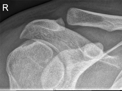
This 61 year old male presented to the Emergency Department with a painful right shoulder following a fall.
There is a grade III separation of the right AC joint.

This patient has a grade III AC joint separation. In addition, the coracoclavicular ligament has avulsed a segment of bone from the inferior aspect of the clavicle. (sometimes referred to as a conoid process or conoid tubercle avulsion fracture)

... back to the Wikiradiograhy home page
... back to the Applied Radiography home page