Abdominal Radiography of the Morbidly Obese Patient
Abdominal radiography of morbidly obese patients introduces unique radiographic challenges. This page examines radiographic techniques that can be employed when you are called upon to provide abdominal radiography of morbidly obese patients.
Definition of Morbidly Obese
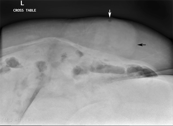
Related Wikiradiography PagesA morbidly obese person is someone who has a body mass index (BMI) over 40. In practical radiographic terms, it is difficult to state a patient weight that will require consideration of special radiographic techniques- radiographic difficulties will depend on the height-to-weight ratio of the patient and the distribution of the weight. As a rule of thumb, patients who weigh over 130kgs (around 300 lbs) will potentially require radiography that takes account of the patients weight . Morbidly obese patients can be referred for abdominal radiography because they are over the weight/size limit for CT scanning.
Manual Handling
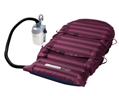
Hovermat
<a class="external" href="http://www.hovermatt.com/main/products/hovermatt/reusable" rel="nofollow" target="_blank">http://www.hovermatt.com/main/products/hovermatt/reusable</a>
<embed allowfullscreen="true" height="350" src="http://widget.wetpaintserv.us/wiki/wikiradiography/widget/youtubevideo/c9b1357a1900e71c86426641ecc568c6cc801507" type="application/x-shockwave-flash" width="425" wmode="transparent"/>If you consider that a safe weight for a radiographer to lift is 20kgs (44 lbs), a patient weighing 150 Kgs (330 lbs) suggests a formidable task in terms of transferring the patient on and off the X-ray table. There are a number of mechanical devices which can provide assistance. Patient lifting cranes can be a device of choice for these patients. The <a class="external" href="http://www.hovermatt.com/main/products/hovermatt/reusable" rel="nofollow" target="_blank">hovermat</a> is also a highly effective device. Slide boards and roll boards also help to reduce friction when sliding patients onto the X-ray table.
Hovermats can be left under obese patients in the intensive care unit. Inflating the hovermat makes the task of inserting an X-ray cassette under a patient much easier.
Radiography
One Person Rollboard Patient Transfer
<embed allowfullscreen="true" height="350" src="http://widget.wetpaintserv.us/wiki/wikiradiography/widget/youtubevideo/2add21cc396980fec4831141c79a9107d6c4645a" type="application/x-shockwave-flash" width="425" wmode="transparent"/>
Two Person Rollboard Patient Transfer
<embed allowfullscreen="true" height="350" src="http://widget.wetpaintserv.us/wiki/wikiradiography/widget/youtubevideo/fd284871b1028ce173083fc04f25a5248c5180e0" type="application/x-shockwave-flash" width="425" wmode="transparent"/>The rollboard renders patient transfers easier by reducing friction. Note that when using a rollboard, the technique is one of pushing the patient not pulling.
There are a number of radiographic technique issues associated with abdominal radiography of morbidly obese patients.
Grid
A grid should be considered essential. Failure to use a grid will result in an image that is degraded by scatter radiation to the point of being undiagnostic.
Cassette Mapping
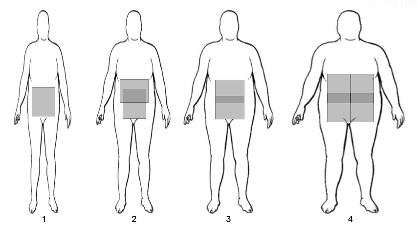
The number of cassettes required and their orientation (portrait or landscape) should be established before any exposure is made. For very large patients, 4 cassettes may be required.
CentringCentring the X-ray beam may not be easy. It has been suggested that the patient's elbow be used as a guide to the level of the patient's iliac crest (source: XrayDude)
Exposure Technique
The kVp selected should be sufficient to penetrate the patient's abdomen- some of the X-ray photons need to make it to the IR! A higher kVp selection will also help to reduce exposure time but will also reduce contrast. Selection of a high mA (within the limits of the X-ray tube and generator) will help to reduce exposure time. Large focal spot size is required.
Table Weight Limit
Manufacturers will provide a safe weight limit for your X-ray table. It is a good idea to label the table with this value for ease of reference.
Pathology Considerations
It is desirable to image the pathology on a single image such that the radiologist (or reporting radiographer) does not need to stitch images together. For example, where a patient has a known or suspected sigmoid volvulus, it is preferable to start with a conventional portrait 35 x 43 cm (17 x 14 inch) position
DR vs CR
DR imaging can provide excellent results when undertaking abdominal radiography of morbidly obese patients and is arguable superior to CR in these patients
Beam Collimation
If you are using a machine that does not automatically collimate the beam to the cassette size, it is well worth pre-collimating to ensure that the beam does not fall outside of the cassette/IR. A round cone is a useful device for scatter reduction with thick anatomy but has little application in abdominal radiography.
Collimation
Mapping
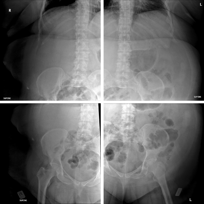
This is a 4 quadrant approach to abdominal radiography. Whilst there is considerable overlap of anatomical areas, the difficulty of achieving a result such as this is easy to underestimate.
Note that the IR shape is square- this is a legacy of using a DR machine which is capable of providing an IR size of 43 x 43 cms (17 x 17 inches).
... back to the Wikiradiography home page
... back to the Applied Radiography page


