Abdominal Plain Film Anatomy

From wikiRadiography
Jump to navigation Jump to search
Introduction

The identification of the anatomy demonstrated on an abdominal plain is variable. It is almost impossible to guarantee the demonstration of abdominal viscera on anay one abdominal plain film. Structures such as the psoas muscle and spleen for example will be demonstrated clearly on some abdominal plain films and completely obscured on others. The reasons can be projectional, position, structural, pathological and chance. This page considers the normal appearances and normal variations in appearances of the normal features demonstrated on abdominal plain film.


The Large Bowel
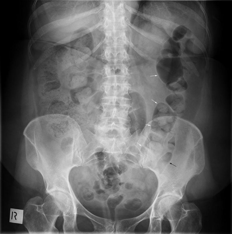
abdomenThe position and course of the large bowel is variable. The orientation of the large bowel is sometimes described as "framing" the abdomen but this is not always the case. This patient has a transverse colon (white arrows) that dips down into the pelvis. This feature is not uncommon and can cause confusion if not appreciated.

One of the features that allows reliable differentiation of the large bowel and small bowel is the presence of faeces in the large bowel. This faeces will usually have a fluid-like consistency as it enters the large bowel from the small bowel, but should have become more solid forming a stool by the time it reaches the splenic flexure. This can be appreciated in this image- the faecal content of the caecum is mottled and uniform compared to the faces in the more distal colon which appears to be formed.

The descending colon (black arrow) is often more ribbon-like, exhibiting less of the characteristic wall features of the transverse colon. The transverse colon is one of the least dependent parts of the large bowel with the patient in the supine position. As such, air in the colon tends to be seen in the transverse colon in supine abdominal plain film radiography- this normal luminal air is a contrast agent and allows the internal wall features to be demonstrated. This air will not only highlight the normal features of the colonic mucosa- it will also highlight abnormal features. This has led to the mistaken notion that bowel disease is commonly sited in the transverse colon.