Abdominal Calcifications
Jump to navigation
Jump to search
Introduction
Gall Stones
Foetus
Ingested Foreign Body
Gall Stones
Abdominal calcifications are commonplace. They can produce confusing appearances and their significance is not always clear. This page considers all aspects of abdominal calcifications as they appear on abdominal plain film.
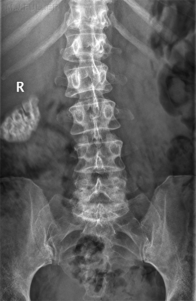
Gall Stones
Foetus
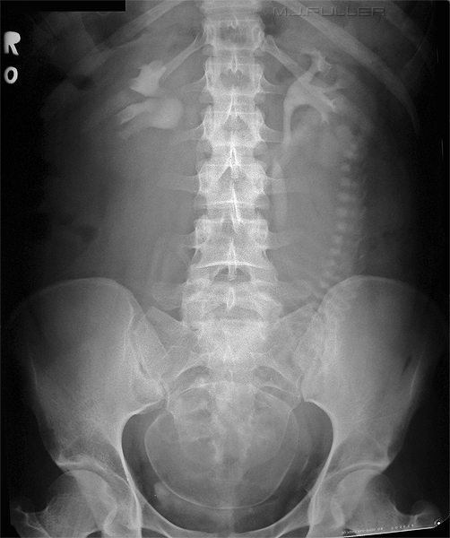
This is a non-pathological calcification- the normal calcification of the foetal skeleton in utero taken from an IVU series.
Ingested Foreign Body
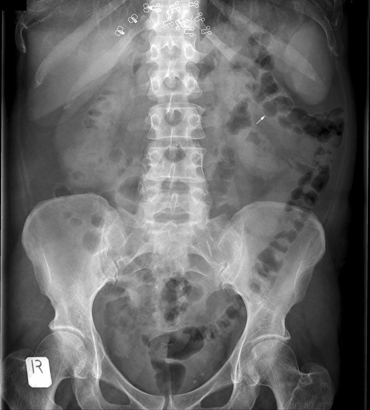
The arrowed structure is an ingested chicken bone which had previosly been demonstrated radiographically on a lateral soft tissue neck image.
Bra hardware noted
Gall Stones
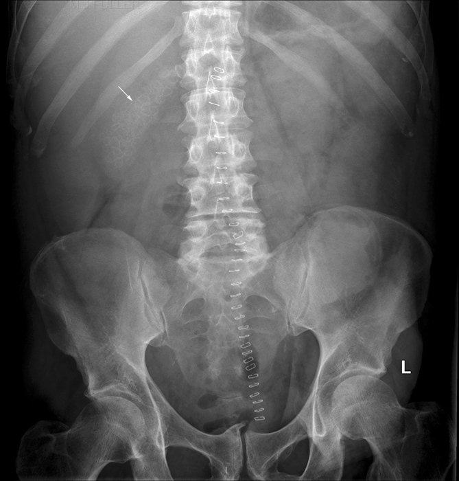
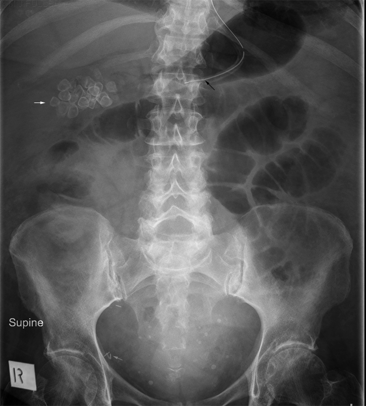
- multiple gallstones (white arrow)
- nasogastric tube (black arrow)
- surgical staples (grey arrow)
- pelvis phleboliths (not marked)
