Abdomen
Jump to navigation
Jump to search
Go Back to Surface Anatomy Index
Abdomen Surface Anatomy | Other pages of interest |
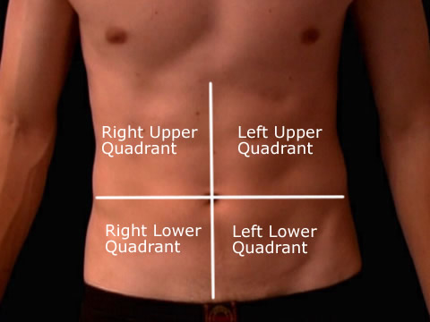
Quadrants of Abdomen
| | Topographically, the abdomen can be divided into right and left upper and right and left lower quadrants by vertical and horizontal lines through the umbilicus. The quadrants are often abreviated on the request form to RUQ, RLQ, LUQ and LLQ. |
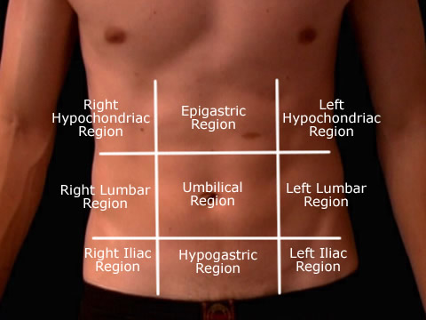
Nine Regions of Abdomen
| | The abdomen may also be divided into nine regions by two longitudinal lines (right and left midclavicular lines) and two transverse planes (subcostal and interspinous planes). The regions are: right and left hypochondriac, right and left lumbar, right and left inguinal (or iliac), epigastric, umbilical and hypogastric. |
Organs of the Abdomen
| The Liver, Spleen and Appendix are common sites of pain in the abdomen. The liver highlighted in red is in the RUQ. The Spleen highlighted in green is in the LUQ. The Appendix in yellow is in the RLQ or more specifically McBurney's Point. |
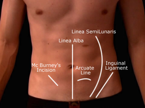
The Abdominal Wall
| | McBurney's point is halfway between the umbilicus and the ASIS and a common location that surgeons use for an incision to remove the appendix. The linea alba is a fibrous structure that runs down the midline of the abdomen and seperates the left and right rectus abdominus muscles. The arcuate line demarcates the lower limit of the posterior layer of the rectus sheath. The inguinal ligament is a band running from the pubic tubercle to the anterior superior iliac spine, its anatomy is very important for operating on hernia patients.The linea semilunaris is a curved tendinous line placed one on either side of the rectus abdominus and corresponds with the lateral border of the rectus muscle. |
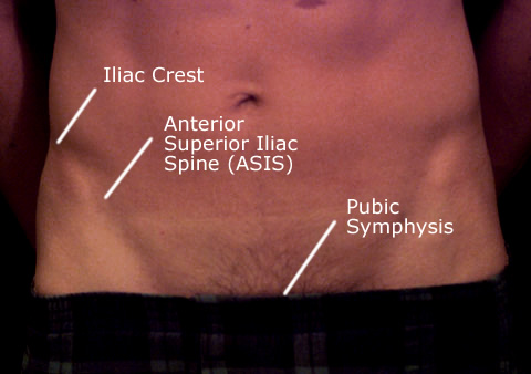
Bony Landmarks of the Lower Abdomen
| | When imaging the abdomen the common bony landmarks used by radiographers are the Iliac crest, ASIS, and the Pubic Symphysis. |
Go Back to Surface Anatomy Index


