Wrist Measurements
RadiographyWhen examining wrist plain film images it is useful to have an understanding of the normal alignment of the wrist. This page considers the normal wrist measurements and how they are utilised.
Comment
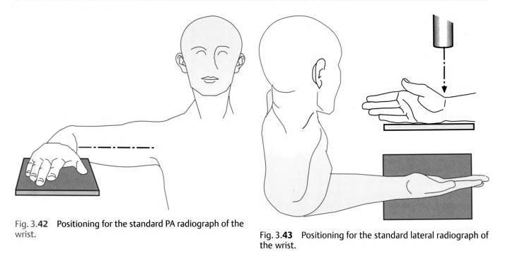
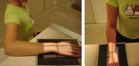
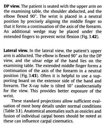
These descriptions are from radiology-based texts rather than radiography texts. The radiography technique textbooks do not consider the position of the forearm/elbow when describing wrist positioning. The positions as described above are akin to 'mixed forearm' views which are performed when patients have limited movement in their arm (usually from pain) or when the patient has an above elbow cast. The wrist positioning approach described above will tend to produce two views of the anatomy at 90 degrees because the relationship between the radius and ulna does not change- the 90 degree movement occurs largely at the shoulder.
adapted from
<a class="external" href="http://books.google.com.au/books?id=G1gWHR1_J9UC&pg=PA54&lpg=PA54&dq=wrist+pronation+supination&source=bl&ots=b7aEg3_V3A&sig=cOaOxDQ3blhQsbSAY_mz-70mSR8&hl=en&ei=BIUaSs7eE4-IkAXrkj0&sa=X&oi=book_result&ct=result&resnum=5#PPA55,M1" rel="nofollow" target="_blank">Raoul Tubiana, Jean-Michel Thomine, Evelyn Mackin</a><a>Examination of the Hand and Wrist. Edition: 2, 1998</a>
<a class="external" href="http://books.google.com.au/books?id=G1gWHR1_J9UC&pg=PA54&lpg=PA54&dq=wrist+pronation+supination&source=bl&ots=b7aEg3_V3A&sig=cOaOxDQ3blhQsbSAY_mz-70mSR8&hl=en&ei=BIUaSs7eE4-IkAXrkj0&sa=X&oi=book_result&ct=result&resnum=5#PPA55,M1" rel="nofollow" target="_blank">Raoul Tubiana, Jean-Michel Thomine, Evelyn Mackin</a>
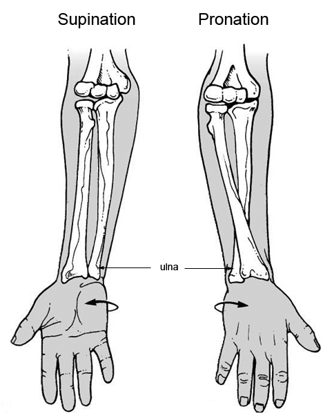
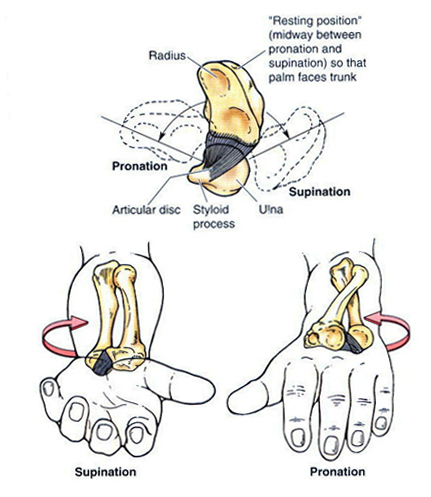
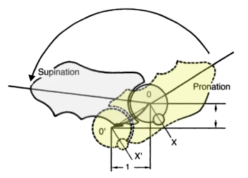
<a class="external" href="http://books.google.com.au/books?id=G1gWHR1_J9UC&pg=PA54&lpg=PA54&dq=wrist+pronation+supination&source=bl&ots=b7aEg3_V3A&sig=cOaOxDQ3blhQsbSAY_mz-70mSR8&hl=en&ei=BIUaSs7eE4-IkAXrkj0&sa=X&oi=book_result&ct=result&resnum=5#PPA55,M1" rel="nofollow" target="_blank"> </a><a><a> </a>Examination of the Hand and Wrist. Edition: 2, 1998</a>The radius undergoes a rotation of almost 180 degrees from full pronation to supination. During this movement of the radius, the ulna translates through a short arc of a circle. This video shows the movement of the distal radius and ulna during the movement of the wrist from PA to lateral. If you watch the ulna (and ulnar styloid), there is no rotation movement.
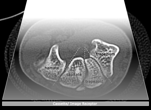
AP/PAThere is a difference between a PA and AP wrist image. The difference is all about the divergence of the X-ray beam. The image below is taken from an axial CT scan of the wrist. Note that the orientation of the carpal bones is radial- they are directed towards a point that is on the volar aspect of the wrist. An AP view of the wrist (as represented below) will tend to benefit from this orientation of the carpal bones by profiling the carpal bones, whereas, in a PA projection, the divergent X-ray beam will work against the radial layout of the carpal bones. The AP projection will tend to be better at demonstrating parallelism of the carpal bones.
Note that this effect is limited. The divergence of the X-ray beam at 115cm is vastly different to the radial-like orientation of the carpal bones as displayed below. An AP projection of the wrist with a short FFD may be of some benefit if carpal alignment/parallelism is of particular interest.
A further technique modification that can be employed to advantage is to flex the patient's fingers to cause the carpus to come into closer contact with the cassette/IR.
Wheeless' Textbook of Orthopaedics,
Posterior Anterior View of the Wrist.
<a class="external" href="http://www.wheelessonline.com/ortho/posterior_anterior_view_of_the_wrist" rel="nofollow" target="_blank">http://www.wheelessonline.com/ortho/posterior_anterior_view_of_the_wrist</a>
LateralA neutral lateral projection of the wrist may be obtained with the arm adducted to the body wall, the elbow flexed 90degrees, the wrist held with no ulnar or radial deviation and no palmar flexion or dorsiflexion.
<a class="external" href="http://radiology.rsnajnls.org/cgi/reprint/184/1/15.pdf" rel="nofollow" target="_blank">F. A. Mann, MD . Anthony J. Wilson, MB, ChB Louis A. Gilula, MD. </a>
<a class="external" href="http://radiology.rsnajnls.org/cgi/reprint/184/1/15.pdf" rel="nofollow" target="_blank">Radiographic Evaluation ofthe Wrist:</a>
<a class="external" href="http://radiology.rsnajnls.org/cgi/reprint/184/1/15.pdf" rel="nofollow" target="_blank">What Does the Hand Surgeon Want to Know?’</a>
<a class="external" href="http://radiology.rsnajnls.org/cgi/reprint/184/1/15.pdf" rel="nofollow" target="_blank">Radiology, Volume 184, Number 1</a>
Wrist/Hand/ForearmYou can't expect to achieve good diagnostic standards with the wrong images. The following is a quote from John Harris on the subject.
"Anatomically and radiographically, the forearm is not the wrist and the wrist is not the hand. Therefore, it is important for attending physicians to indicate precisely the anatomical part to be examined radiographically. Specifically, the attending physician must define, on the basis of history and physical examination, whether the forearm, wrist, or hand, is to be examined, since the routine views of one of these anatomic areas is of limited practical value with respect to the others. In spite of this admonition, it is important to remember that injuries of the distal forearm may cause, or be associated with, carpal injuries. Finally, contrary to an often-repeated dictum, it is not necessary to radiographically examine both the wrist and the elbow in the instance of a suspected radial or ulnar shaft fracture in a patient who is alert, responsive, and communicative and on whom a proper physical examination can be performed. In patients who do not fulfil these criteria, it is prudent to examine both the wrist and the elbow." John Harris, William Harris, Robert Novelline.
The Radiology of Emergency Medicine, Third edition.
Williams and Wilkins, 1993
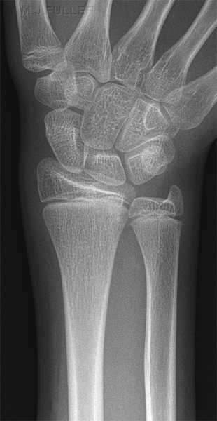
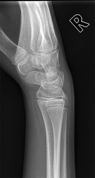
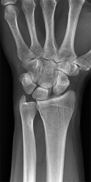
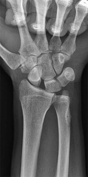
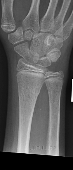
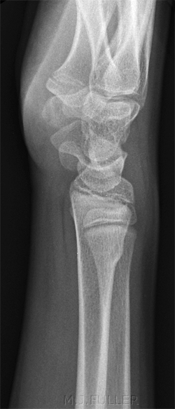
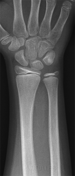
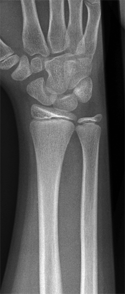
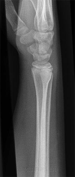
These two images of a child's wrist demonstrate that the ulna is in the same position for both projections. If there was a fracture of the ulna (in addition to the fracture of the radius), it could easily be missed because there is only a single projection of the ulna.
To permit pronation and supination, the head of the ulna rotates in the sigmoid notch of the radius. <a class="external" href="http://books.google.com.au/books?id=qTCbB2N2UFcC&pg=PA54&lpg=PA54&dq=disi+visi&source=bl&ots=jrX-iBQZ9o&sig=ueTw6hUN5wk__z_SNTKm_3X0V6A&hl=en&ei=ikgTSvaNCJS-tAOy68XdDQ&sa=X&oi=book_result&ct=result&resnum=4#PPA55,M1" rel="nofollow" target="_blank">Giuseppe Guglielmi, Cornelis Van Kuijk, Harry K. Genant, Fundamentals of hand and wrist imaging, 2001</a> If this was not the case, the images to the left would not be possible- that is, the radius could not be imaged in two views at 90 degrees without the ulna similarly being imaged at right angles.
Rotation in the distal radioulnar joint (DRUJ) takes place around the ulna as a fixed point around which the radius rotates along with the hand.<a class="external" href="http://books.google.com.au/books?id=67UjC6sFy04C&pg=PA120&lpg=PA120&dq=meniscus+homologue&source=bl&ots=LbhEMWnz-M&sig=yf1l2Js5wqLx8XAzBncGuRnz6Ds&hl=en&ei=D8ETSp6pKtGUkAW0p4T9Dg&sa=X&oi=book_result&ct=result&resnum=11#PPA125,M1" rel="nofollow" target="_blank">Ulrich Lanz, Rainer Schmitt, Wolfgang Buchberger, Diagnostic Imaging of the Hand, 2008</a>This explains why you can rotate the wrist from a PA into a lateral position without the ulna moving (see left). It also explains why you shouldn't execute this manoeuvre when changing from a PA wrist radiographic position to a lateral wrist position- the change should involve movement of the whole forearm.
Anatomy
You tend to remember those things that you regularly use. For example, the vast majority of radiographers will be able to identify the scaphoid on a PA/AP wrist image.
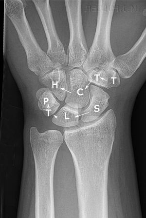
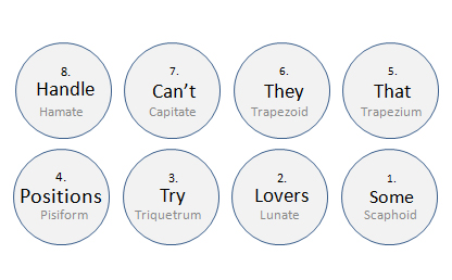
There's nothing like a mnemonic for remembering those things that you don't use very often. One of the most common and useful mnemonic types is the acronym. It seems that the easiest acronyms to remember are the risqué ones and this one is no exception. The carpal bones can be thought of as two rows of 4 bones. The memory aid acronym is as followsSome Lovers Try Positions That They Can't Handle.
Read in number order.A few other acronyms for the carpal bones
•Sally Left The PartyTo Take Cathy Home
•She Looks Too PrettyTry To Catch Her
- Some Ladies Try Painting
Their Nails Curious Hues
- So Long to Pinky
Here Comes the Thumb (clever!)
"Another mnemonic, useful for remembering the order of the trapezium and trapezoid, is that "trapezium" rhymes with "thumb" and articulates with it."
<a class="external" href="http://www.hand-therapy.com/images/Radiology_100500.pdf" rel="nofollow" target="_blank">Karen Nugent, PT, CHT and David Nelson, M.D. RADIOLOGY OF THE WRIST</a>If you are one of those wordsmiths that like to know the origins/derivations of words, you may find the following helpfulScaphoid
- Greek origins, boat shaped, "skaphe" might be better translated as "skiff".
Lunate
- lunar shaped (half moon) on lateral image and trapezoidal on PA
Triquetrum
- as the name suggests, it is triangular
Pisiform
- pea shaped, the only carpal sesamoid bone
Trapezium
- table shaped
Trapezoid
- same origins as trapezium- table shaped
Capitate
- head (capit - head)
Hamate
- hooked. ( Latin hamatus "hooked," from hamus which means "hook.")
<a class="external" href="http://www.hand-therapy.com/images/Radiology_100500.pdf" rel="nofollow" target="_blank">Karen Nugent, PT, CHT and David Nelson, M.D. RADIOLOGY OF THE WRIST</a>
Alignment and Measurement
Parallelism
<embed allowfullscreen="true" height="350" src="http://widget.wetpaintserv.us/wiki/wikiradiography/widget/youtubevideo/907c53b0de96f656eb00ac0d8b3c92b88bd3558c" type="application/x-shockwave-flash" width="425" wmode="transparent"/>
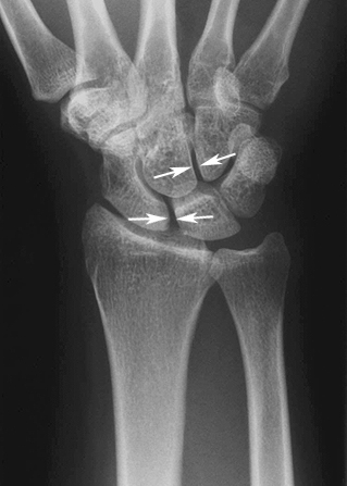
(Note: Youtube videos are displayed at lower resolution when 'hot-linked'. Click on the bottom right corner of the video and it will open at full resolution.)All of the carpal bones have approximately the same thickness of articular cartilage and normally lie in apposition to the adjacent carpal bone. In other words, normally there is no space between cartilage surfaces of adjacent carpal bones. It follows that the joint spaces seen on the PA view should be approximately the same width.
<a class="external" href="http://www.hand-therapy.com/images/Radiology_100500.pdf" rel="nofollow" target="_blank">RADIOLOGY OF THE WRIST
Presented by Karen Nugent, PT, CHT and David Nelson, M.D. At the 23rd Annual Meeting of the American Society of Hand Therapists
5 October 2000</a>
Normally the intercarpal, carpometacarpal and radiocarpal joint spaces are 2mm wide or less. When a joint is > 4mm wide it is usually abnormal. Parallelism can only be applied to the joints that are demonstrated in profile. (Gilula, L.A. and Totty W.G. The Traumatised Hand and Wrist, Radiographic and Anatomic Correlation, 1992, p221).
The MRI wrist demonstrates that the intercarpal distances are approximately equal
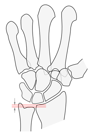
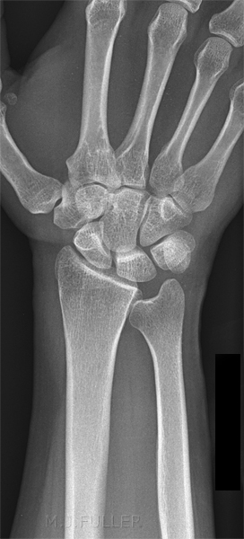
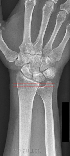
Ulnar Variance syn. radioulnar index
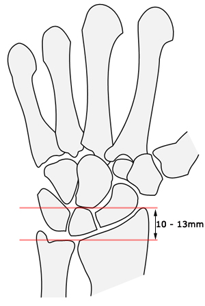
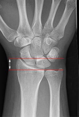
Radial length or Radial height
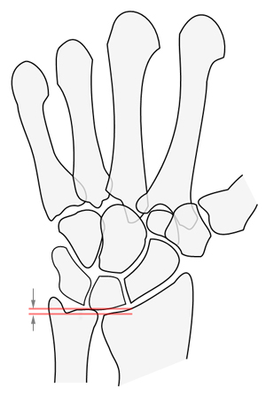
Ulnar variance refers to the position of the cortical margin of the distal ulna relative to that of the distal radius. It gives insight into the degree of axial loading transmitted to the lunate and may therefore be helpful in determining the aetiology of repetitive wrist trauma. Medscape General Medicine, 1999, Imaging of the Wrist and Hand,
<a class="external" href="http://cme.medscape.com/viewarticle/408495_2" rel="nofollow" target="_blank">http://cme.medscape.com/viewarticle/408495_2</a>
Ulnar variance is defined as neutral, positive (plus), or negative (minus) on the basis of whether the distal articular surface of the ulna is aligned with the distal articular surface of the radius on a neutral posteroanterior radiograph, or lies distal (ulnar plus) or proximal (ulnar minus) to the radius (4-6).
All patients with fractures of either of the forearm bones require careful evaluation of both wrist and elbow, and special attention should be given to the distal radioulnar joint and ulnar variance.
<a class="external" href="http://radiology.rsnajnls.org/cgi/reprint/184/1/15.pdf" rel="nofollow" target="_blank">F. A. Mann, MD . Anthony J. Wilson, MB, ChB Louis A. Gilula, MD.
Radiographic Evaluation ofthe Wrist:
What Does the Hand Surgeon Want to Know?’
Radiology, Volume 184, Number 1</a>
Ulnar variance is independent of the length of the ulnar styloid process, which may also vary. However, wrist position is an important determinant of ulnar variance (2,6 –9). Maximum forearm pronation results in an increase in positive ulnar variance, whereas maximum forearm supination decreases ulnar variance. Ulnar variance increases significantly with a firm grip and
returns to its original state with cessation of grip.
<a class="external" href="http://radiographics.rsnajnls.org/cgi/reprint/22/1/105?maxtoshow=&HITS=10&hits=10&RESULTFORMAT=&fulltext=wrist+X-ray&searchid=1&FIRSTINDEX=0&sortspec=relevance&resourcetype=HWCIT" rel="nofollow" target="_blank">Luis Cerezal, MD ● Francisco del Pinal, MD, PhD ● Faustino Abascal, MD ● Roberto Garcı´a-Valtuille, MD ● Teresa Pereda, MD ● Ana Canga, MD
Imaging Findings in Ulnar-sided Wrist Impaction Syndromes. RadioGraphics 2002; 22:105–121</a>
Radial Inclination syn Radial Angle, Radial inclination, radial deviation
Radial length or height
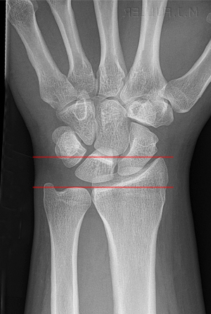
Radial length is measured on the PA radiograph as the distance between one line perpendicular to the long axis of the radius passing through the distal tip of the radial styloid.
A second line intersects distal articular surface of ulnar head. This measurement averages 10-13 mm.
quoted from <a class="external" href="http://www.radiologyassistant.nl/en/476a23436683b" rel="nofollow" target="_blank">http://www.radiologyassistant.nl/en/476a23436683b</a>
Loss of radial height is a predictor of a less favourable outcome in distal radial fractures. The normal mean has been reported to be 11-12 mm (26). (also reported as 13.5 mm ± 3.8 (2 standard deviations)
<a class="external" href="http://radiology.rsnajnls.org/cgi/reprint/184/1/15.pdf" rel="nofollow" target="_blank">F. A. Mann, MD . Anthony J. Wilson, MB, ChB Louis A. Gilula, MD.
Radiographic Evaluation ofthe Wrist:
What Does the Hand Surgeon Want to Know?’
Radiology, Volume 184, Number 1</a>
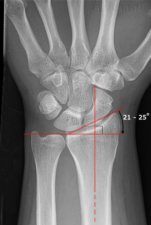
Radiocarpal Angle
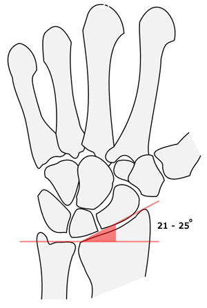
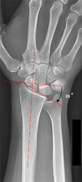
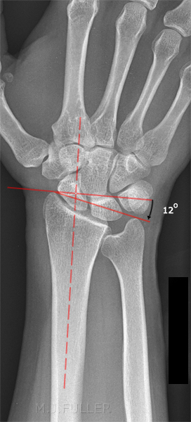
Radial inclination
Radial inclination represents the angle between one line connecting the radial styloid tip and the ulnar aspect of the distal radius and a second line perpendicular to the longitudinal axis of the radius.
The radial inclination ranges between 21° and 25°. (another reference quotes 16- 28 degrees). Normal is reported as 25.4 degrees with a standard deviation of 2.2 degrees.
Loss of radial inclination will increase the load across the lunate.
quoted from <a class="external" href="http://www.radiologyassistant.nl/en/476a23436683b" rel="nofollow" target="_blank">http://www.radiologyassistant.nl/en/476a23436683b</a>
Patients treated for distal radial fractures who have a radial inclination of less than 5 degrees have poorer results than those with normal or near normal inclination.
<a class="external" href="http://radiology.rsnajnls.org/cgi/reprint/184/1/15.pdf" rel="nofollow" target="_blank">F. A. Mann, MD . Anthony J. Wilson, MB, ChB Louis A. Gilula, MD.
Radiographic Evaluation ofthe Wrist:
What Does the Hand Surgeon Want to Know?’
Radiology, Volume 184, Number 1</a>
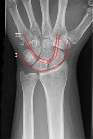
The Carpal Arcs syn. Gilula's arcs
Carpal Height
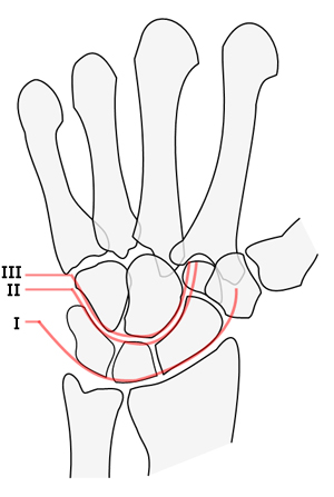
The concept of three radiographic arcs was first proposed by Gilula in 1979. The carpal bones all have rounded edges to varying degrees, and as such the arcs have small indentations at the joint lines. However, there should be no stepoffs in the contour of the arcs. Such a "broken arc" implies a ligament tear or fracture at the site of the broken arc.
<a class="external" href="http://www.hand-therapy.com/images/Radiology_100500.pdf" rel="nofollow" target="_blank">
</a>
Three carpal arcs: smooth curves joining the surfaces of the carpal bones as shown on the left.
Carpal Arc I
The first arc is a smooth curve outlining the proximal convexities of the scaphoid, lunate and triquetrum.
Carpal Arc II
The second arc traces the distal concave surfaces of the same bones
Carpal Arc III
The third arc follows the main proximal curvatures of the capitate and hamate.
adapted from
<a class="external" href="http://www.radiologyassistant.nl/en/42a29ec06b9e8" rel="nofollow" target="_blank">http://www.radiologyassistant.nl/en/42a29ec06b9e8</a>
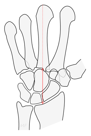
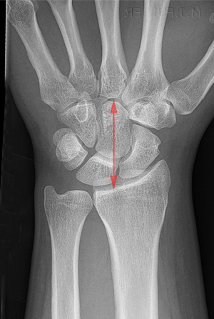
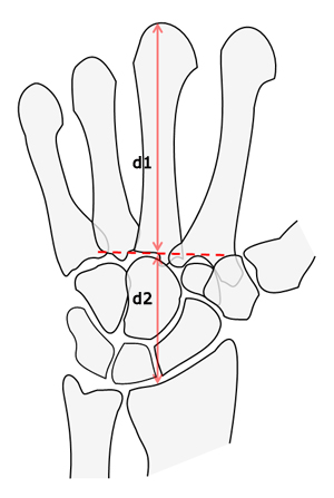
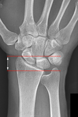
Carpal Height Ratio
"Carpal height is a radiologic concept to aid in the quantification of carpal collapse. Sequential measurements can aid in the assessment of
disease severity and progression. Carpal height is defined as the distance from the base of the third metacarpal to the subchondral sclerotic line of the distal radial articular surface as measured
along the axis extended from the third metacarpal."<a class="external" href="http://radiology.rsnajnls.org/cgi/reprint/184/1/15.pdf" rel="nofollow" target="_blank">F. A. Mann, MD . Anthony J. Wilson, MB, ChB Louis A. Gilula, MD. </a>
<a class="external" href="http://radiology.rsnajnls.org/cgi/reprint/184/1/15.pdf" rel="nofollow" target="_blank">Radiographic Evaluation ofthe Wrist:</a>
<a class="external" href="http://radiology.rsnajnls.org/cgi/reprint/184/1/15.pdf" rel="nofollow" target="_blank">What Does the Hand Surgeon Want to Know?’</a>
<a class="external" href="http://radiology.rsnajnls.org/cgi/reprint/184/1/15.pdf" rel="nofollow" target="_blank">Radiology, Volume 184, Number 1</a>
The radiographic technique for carpal height measurement requires that the hand be positioned for a neutral posteroanterior examination. Specifically, the shoulder should be abducted 90 degrees, elbow flexed 90 degrees, the wrist without ulnar or radial deviation and without palmar flexion or dorsiflexion.<a class="external" href="http://radiology.rsnajnls.org/cgi/reprint/184/1/15.pdf" rel="nofollow" target="_blank">F. A. Mann, MD . Anthony J. Wilson, MB, ChB Louis A. Gilula, MD. </a>
<a class="external" href="http://radiology.rsnajnls.org/cgi/reprint/184/1/15.pdf" rel="nofollow" target="_blank">Radiographic Evaluation ofthe Wrist:</a>
<a class="external" href="http://radiology.rsnajnls.org/cgi/reprint/184/1/15.pdf" rel="nofollow" target="_blank">What Does the Hand Surgeon Want to Know?’</a>
<a class="external" href="http://radiology.rsnajnls.org/cgi/reprint/184/1/15.pdf" rel="nofollow" target="_blank">Radiology, Volume 184, Number 1</a>
"The carpal height ratio is usually obtained by dividing the carpal height by the length of the third
metacarpal"
McMurtry (1) found that wrist examinations are often performed without inclusion of all the metacarpals.
He therefore developed an alternative
method for determining the carpal
height ratio by using only the carpal
bones, metacarpal bases, and distal
radius. To calculate this alternative
carpal height ratio, the carpal height,
as defined above, should be divided
by the capitate length.
<a class="external" href="http://radiology.rsnajnls.org/cgi/reprint/184/1/15.pdf" rel="nofollow" target="_blank">F. A. Mann, MD . Anthony J. Wilson, MB, ChB Louis A. Gilula, MD.
Radiographic Evaluation ofthe Wrist:
What Does the Hand Surgeon Want to Know?’
Radiology, Volume 184, Number 1</a>
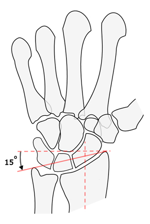
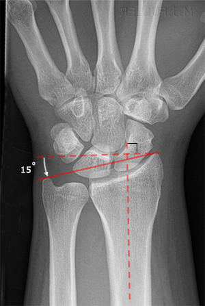
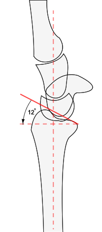
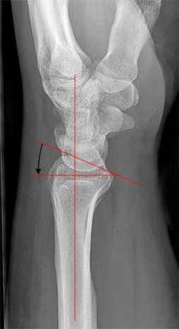
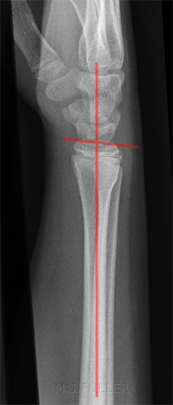
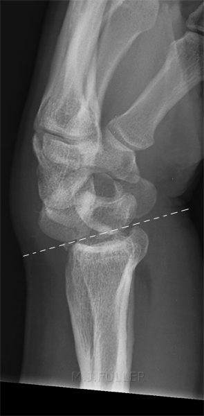
Palmar Tilt syn volar tilt, volar inclination, and palmar slope
Its most common use is in assessing initial and residual deformities associated with fractures of the distal radius and in planning operative correction of disabling deformities. Palmar tilt represents the saggital plane inclination of the distal radial articular surface.
In more than half of the patients with greater than 15 degrees of dorsal angulation of palmar tilt, the result is unsatisfactory in that grip strength and endurance are decreased
Normal measurements vary by report and gender of the subject. Negative values for palmar tilt are not normal. Normal variations have been described as between 0 and 22 degrees with a mean of 14.5 degrees and a standard deviation of 4.3 degrees. Women have been reported to have a palmar tilt of 11.3 - 13.5 degrees with a mean of 12.4 degrees. The same study reports palmar tilt in men to vary between 8.3 degrees and 10.3 degrees.
quoted from
<a class="external" href="http://radiology.rsnajnls.org/cgi/reprint/184/1/15.pdf" rel="nofollow" target="_blank">F. A. Mann, MD . Anthony J. Wilson, MB, ChB Louis A. Gilula, MD.
Radiographic Evaluation ofthe Wrist:
What Does the Hand Surgeon Want to Know?’
Radiology, Volume 184, Number 1</a>
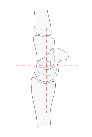
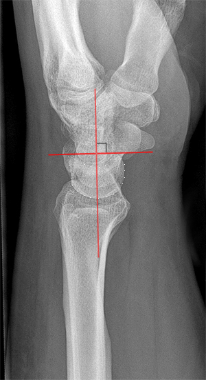
Scaphoid Axis
Lunate Axis
"The true axis of the scaphoid is the line through the midpoints of its proximal and distal poles. Since the midpoint of the proximal pole is often difficult to appreciate, an almost parallel line can be used that is traced along the most ventral points of the proximal and distal poles of the bone"
quoted from<a class="external" href="http://www.radiologyassistant.nl/en/42a29ec06b9e8" rel="nofollow" target="_blank">Louis A. Gilula and Ileana Chesaru</a>
<a class="external" href="http://www.radiologyassistant.nl/en/42a29ec06b9e8" rel="nofollow" target="_blank">Wrist - Carpal instability
The Radiology Assistant</a>
"The axis of the lunate runs through the midpoints of the convex proximal and concave distal joint surfaces and can best be drawn by finding the perpendicular to a line joining the distal palmar and dorsal borders of the bone as demonstrated on the left."
quoted from<a class="external" href="http://www.radiologyassistant.nl/en/42a29ec06b9e8" rel="nofollow" target="_blank">Louis A. Gilula and Ileana Chesaru</a>
<a class="external" href="http://www.radiologyassistant.nl/en/42a29ec06b9e8" rel="nofollow" target="_blank">Wrist - Carpal instability
The Radiology Assistant</a>
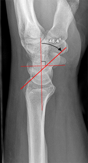
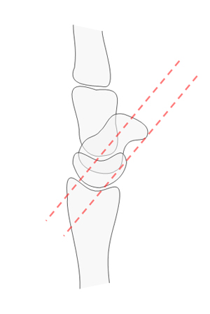
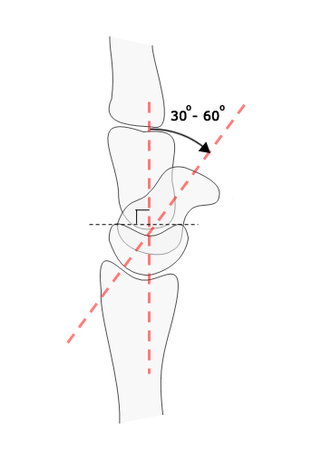
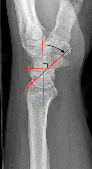
Scapholunate Angle
Scapholunate angle
- Normal: 30 - 60°
- Questionably abnormal: 60 - 80°
- Abnormal: > 80° This indicates instability of the wrist.
quoted from<a class="external" href="http://www.radiologyassistant.nl/en/42a29ec06b9e8" rel="nofollow" target="_blank">Louis A. Gilula and Ileana Chesaru</a>
<a class="external" href="http://www.radiologyassistant.nl/en/42a29ec06b9e8" rel="nofollow" target="_blank">Wrist - Carpal instability
The Radiology Assistant</a>
The numbers are soft, but this is a guide
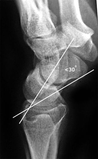
- VISI < 30 degrees
- Normal 30 - 60 degrees
- DISI > 60 degrees
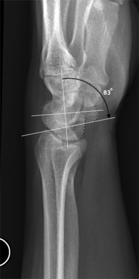
DISI (dorsal intercalated segmental instability)
note that this angle has a larger than normal error because the wrist is not in a true lateral positionDISI, or dorsiflexion instability, is short for
dorsal intercalated segmental instability.
The intercalated segment is the proximal carpal row identified by the lunate. The term 'intercalated segment' refers to it being the part in between the proximal segment of the wrist consisting of the radius and the ulna and the distal segment, represented by the distal carpal row and the metacarpals.
So all this means is that in DISI, or dorsiflexion instability, the lunate is angulated dorsally.
If you think the lunate is tilted, measure the scapholunate angle
(30-60°is normal, 60-80°is questionably abnormal, >80° is abnormal) and the capitolunate angle (<30° is normal).
quoted from<a class="external" href="http://www.radiologyassistant.nl/en/42a29ec06b9e8" rel="nofollow" target="_blank">Louis A. Gilula and Ileana Chesaru</a>
<a class="external" href="http://www.radiologyassistant.nl/en/42a29ec06b9e8" rel="nofollow" target="_blank">Wrist - Carpal instability
The Radiology Assistant</a>
DISI is due to disruption of the scapho-lunate articulation
A DISI will typically demonstrate a scapho-lunate angle greater than 60 degrees
quoted from
<a class="external" href="http://books.google.com.au/books?id=qTCbB2N2UFcC&pg=PA54&lpg=PA54&dq=disi+visi&source=bl&ots=jrX-iBQZ9o&sig=ueTw6hUN5wk__z_SNTKm_3X0V6A&hl=en&ei=ikgTSvaNCJS-tAOy68XdDQ&sa=X&oi=book_result&ct=result&resnum=4#PPA55,M1" rel="nofollow" target="_blank">Giuseppe Guglielmi, Cornelis Van Kuijk, Harry K. Genant, Fundamentals of hand and wrist imaging, 2001</a>
DISI pattern:adapted from <a class="external" href="http://som.flinders.edu.au/FUSA/ORTHOWEB/notebook/regional/wrist.html" rel="nofollow" target="_blank">http://som.flinders.edu.au/FUSA/ORTHOWEB/notebook/regional/wrist.html</a>
- when the scapholunate joint is dissociated, the scaphoid is palmar flexed and the lunate is dorsiflexed
- Scapho-lunate angle usually 30- 60degrees (average 46 degrees) and with DISI it is greater than 70degrees
VISI (Volar intercalated segmental instability)
VISIadapted from
<a class="external" href="http://imaging.birjournals.org/cgi/content/full/15/4/180" rel="nofollow" target="_blank">P S McAlinden and J Teh,
</a><a class="external" href="http://imaging.birjournals.org/cgi/content/full/15/4/180" rel="nofollow" target="_blank">Imaging of the wrist, Imaging 15:180-192 (2003)</a>
While most DISI is abnormal, in many cases VISI is a normal variant, especially if the wrist is very lax.
quoted from<a class="external" href="http://www.radiologyassistant.nl/en/42a29ec06b9e8" rel="nofollow" target="_blank">Louis A. Gilula and Ileana Chesaru</a>
<a class="external" href="http://www.radiologyassistant.nl/en/42a29ec06b9e8" rel="nofollow" target="_blank">Wrist - Carpal instability
The Radiology Assistant</a>
VISI is secondary to disruption of the lunate and triquetral
quoted from
<a class="external" href="http://books.google.com.au/books?id=qTCbB2N2UFcC&pg=PA54&lpg=PA54&dq=disi+visi&source=bl&ots=jrX-iBQZ9o&sig=ueTw6hUN5wk__z_SNTKm_3X0V6A&hl=en&ei=ikgTSvaNCJS-tAOy68XdDQ&sa=X&oi=book_result&ct=result&resnum=4#PPA55,M1" rel="nofollow" target="_blank">Giuseppe Guglielmi, Cornelis Van Kuijk, Harry K. Genant, Fundamentals of hand and wrist imaging, 2001</a>
VISI pattern:adapted from <a class="external" href="http://som.flinders.edu.au/FUSA/ORTHOWEB/notebook/regional/wrist.html" rel="nofollow" target="_blank">http://som.flinders.edu.au/FUSA/ORTHOWEB/notebook/regional/wrist.html</a>
- lunate palmar flexed
- if the lunate and triquetrum can be seen, the normal lunotriquetral angle of approximately -16 degrees becomes neutral or positive
Case 1
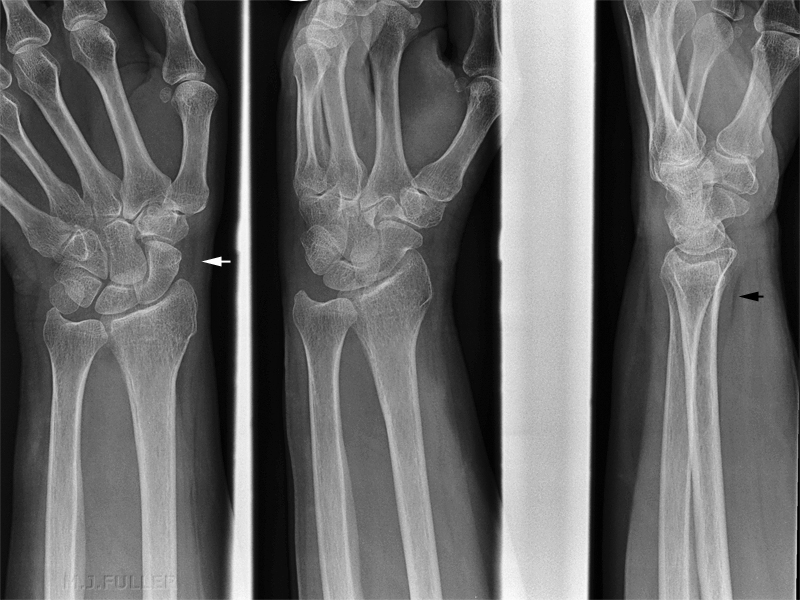
This patient presented to the Emergency Department following a fall. Routine wrist views were performed
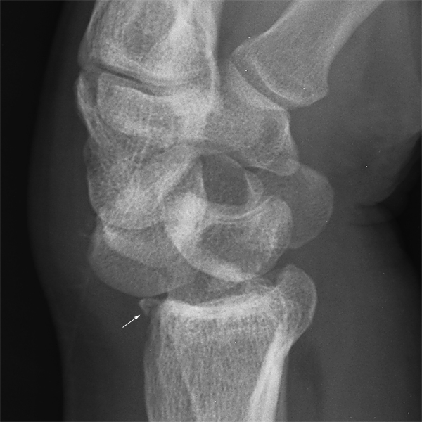
This series demonstrated no obvious fracture. Moreover, the scaphoid fat pad (white arrow) and the pronator fat pad (black arrow) were normal.
The palmar inclination is zero. This is not diagnostic of a fracture, but is suspicious.
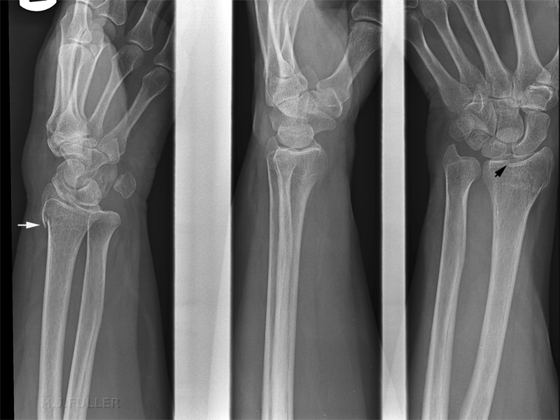
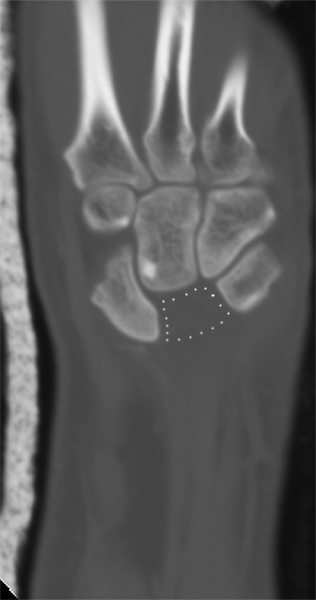
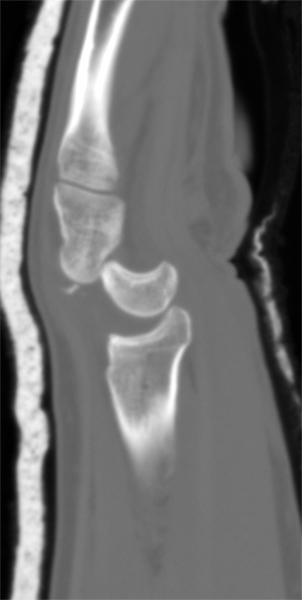
The radiographer was convinced clinically that the patient had sustained a fracture and considered whether the soft tissue signs were misleading (as there are known to be on occasions). Supplementary views were performed as shown below.
Case 2
This13 year old boy presented to the Emergency Department after a fall onto an outstretched hand.
Case 3Comment
The subtle fracture of the wrist is evident from the buckle fracture and supported by the negative palmar tilt.
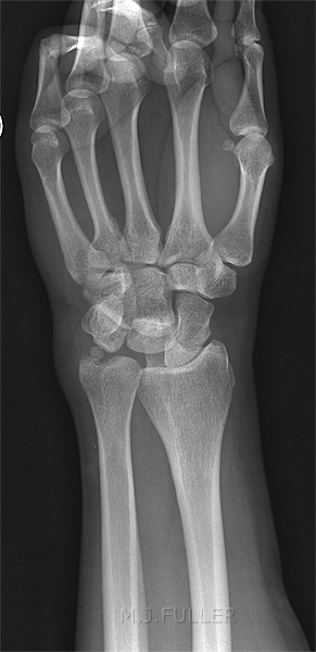
This 34 year old man presented to the Emergency Department after hand vs ceiling fan trauma. The patient has a normal anatomical variant known as short ulna.
The radial height appears to be increased (unable to measure) Normal radial height Comment
The abnormal wrist measurements are a normal anatomical variant known as short ulna.
Case 4
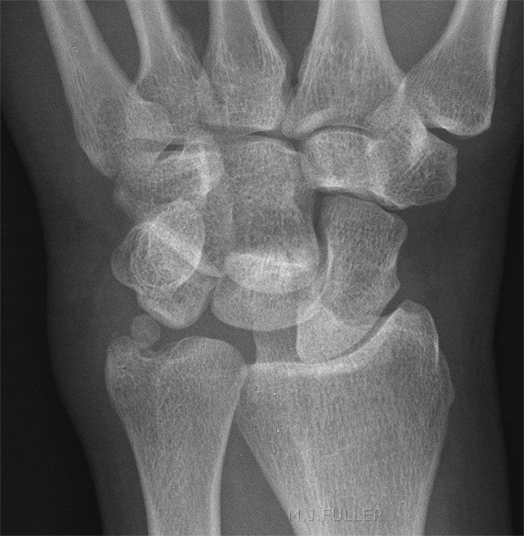
This 32 year old man presented to the Emergency Department after falling through a roof onto a concrete floor.
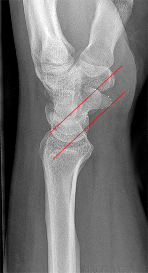
There is loss of parallelism of the carpal bones There is loss of the normal proximal and distal carpal arc lines
DiscussionThe loss of normal carpal arcs was noted by the radiographer who proceeded to perform a lateral wrist view.
The measurements described above cannot be accurately determined without correctly centred and positioned anatomy. Importantly, positioning the patient for forearm imaging (rather than wrist) comes at a high price- if wrist measurements are required forearm views will not suffice.
...back to the Applied Radiography home page
