Introduction This page considers all aspects of urethrography in both acute and non-acute settings.
Case 1  | This 50 year old male presented to the ED by ambulance following a motor vehicle accident.
A radiographic trauma series was undertaken in the resus room. The trauma series revealed several fractures including pelvic fractures.
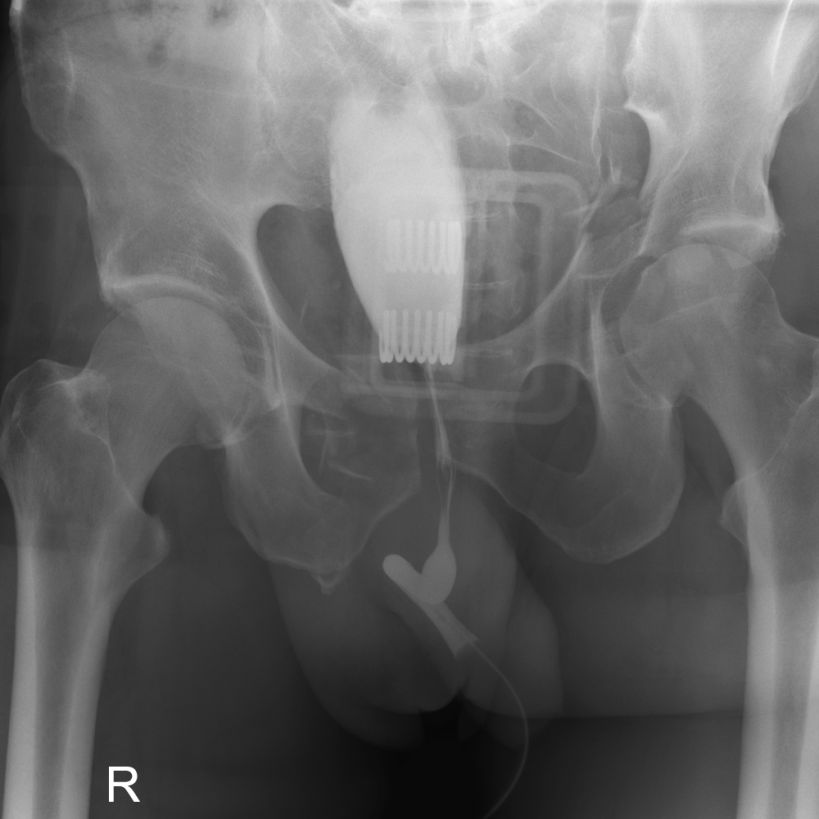
The patient underwent urethrography to asses the urethra for potential traumatic injury.
A Carestream 35 x 43cm DRX cassette was placed in landscape orientation under the patient's pelvis. Contrast injection into the urethra was performed via a foley catheter.
The AP projection does not demonstrate the urethra well. |
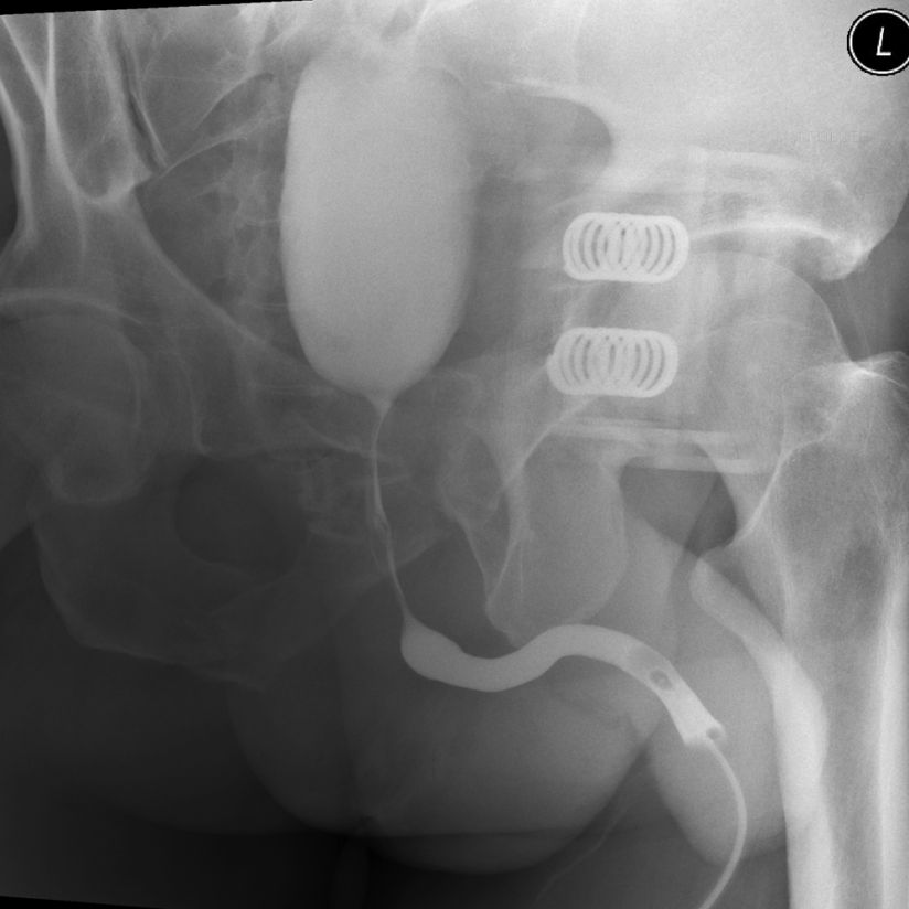
 | The DRX cassette was not moved. The beam was angled across the patient to achieve an LPO projection. The urethra is demonstrated well. |
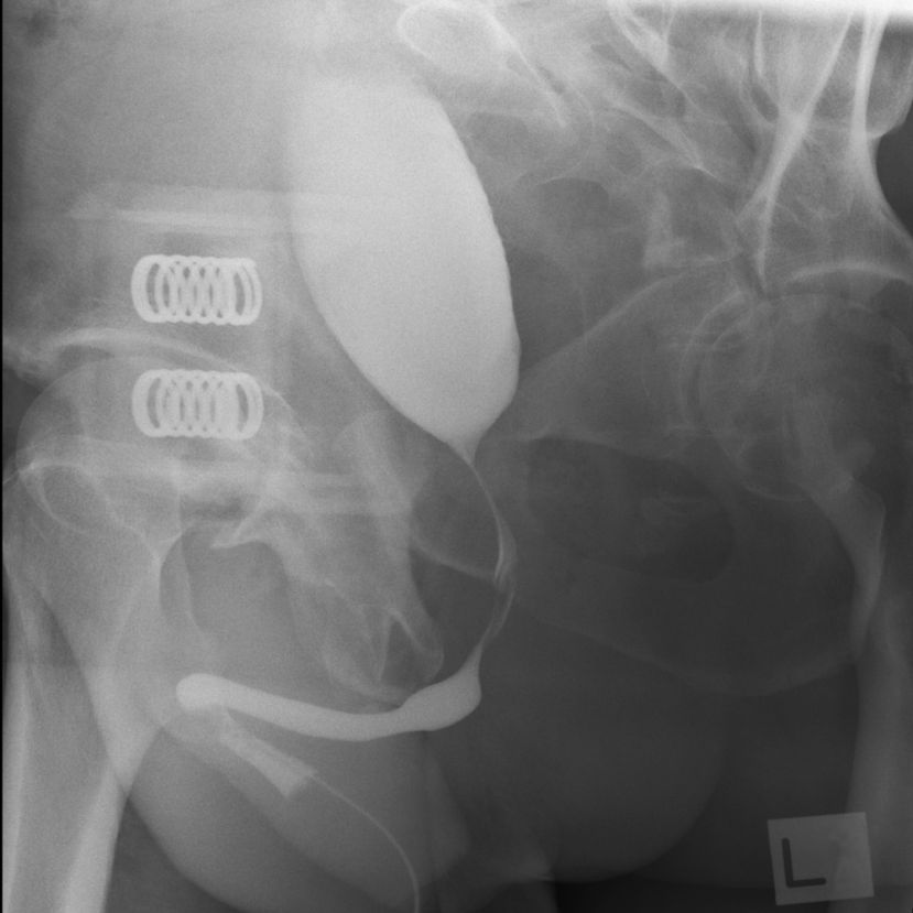
 | The opposite oblique was again achieved without moving the patient or imaging cassette. The urethra appears to be undamaged. The urinary bladder has an abnormal elongated shape associated with pressure from traumatic pelvic haematoma. |
|
|
... back to
wikiradiography home page
... back to
applied radiography


