Trigeminal Nerve Block
Jump to navigation
Jump to search
Introduction
This page details the radiographic technique associated with a trigeminal nerve block procedure. A trigeminal nerve block involves the injection of glycerol into the trigeminal nerve cistern to alleviate trigeminal neuralgia. This is achieved under fluoroscopic control.
Preparation
- Patient Identification (3 C's- correct patient, correct side, correct procedure)
- Is the patient pregnant?
- Is the recovery area expecting the patient? Is the patient intending to drive home after the procedure?
- Change patient into hospital gown if required
- Check departmental policy re signed consent by the patient to the procedure
- Ensure old imaging is on-hand/available
Indications for Trigeminal Nerve Block
- trigeminal neuralgia
Contraindications for Shoulder Arthrography
- recent cervical spine surgery (neck extension contraindicated)
- contrast media or iodine allergy
Anatomy
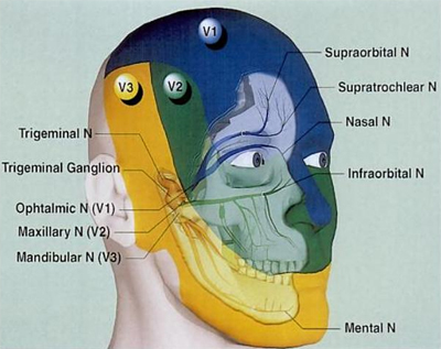
"Neuralgia is the medical term used to describe nerve pain. Trigeminal neuralgia is an extremely painful condition that affects the trigeminal nerve in the face, which is also called the fifth cranial nerve. The trigeminal nerve plays a very important role in the face, being responsible for sensing touch, pressure, pain and temperature in the jaw, gums, forehead and around the sensitive eye area. Since it controls sensation in almost the entire face, pain in the trigeminal nerve can affect many different parts of the face."
<a class="external" href="http://www.irishhealth.com/article.html?con=496" rel="nofollow" target="_blank">www.irishhealth.com</a>
The trigeminal nerve has three main divisions
- Ophthalmic Nerve (V1)
- Maxillary Nerve (V2)
- Mandibular Nerve (V3)
Radiographic Technique
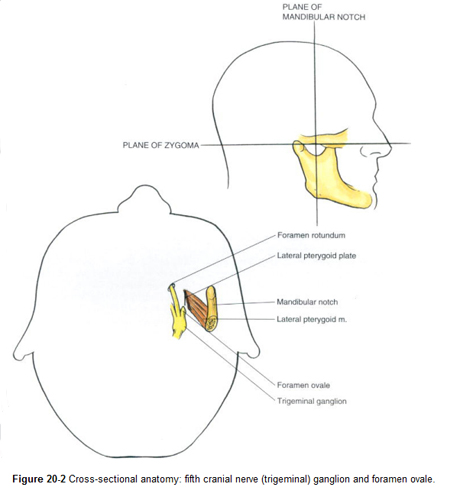
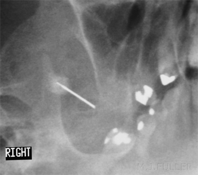
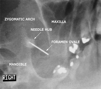
<a class="external" href="http://polanest.webd.pl/pliki/varia/books/AtRegAn/micro189.lib3.hawaii.edu_3a2127/das/book/body/0/1353/i4-u1.0-b1-4160-2239-2..50024-2--f2.fig.htm" rel="nofollow" target="_blank"> Brown: Atlas of Regional Anesthesia, 3rd ed</a>The radiographer's primary role is to demonstrate the foramen ovale using the C-arm image intensifier. Clearly, a sound knowledge of the location of the foramen ovale and the technique to demonstrate it is required.
The patient is positioned supine on the fluoroscopy table with the head in a reverse occipitomental (OM) position (ie chin up/neck extended). The patient's head is rotated 30 degrees away from the affected side with 30 degrees of caudal angulation of the X-ray beam. A C-arm fluoroscopy unit is required to achieve this position.
Small focal spot should be selected and a small field size is desirable.
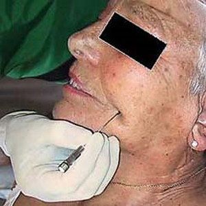
source : <a class="external" href="http://www.schmerzzentrum.ch/infos_zahnaerzte/trigeminusneuralgie.php" rel="nofollow" target="_blank">http://www.schmerzzentrum.ch/infos_zahnaerzte/trigeminusneuralgie.php</a>Whilst maintaining this oblique position, the operator (radiologist/neurosurgeon) cleans the affected cheek with betadine (or similar antiseptic) and then injects local anaesthetic. A needle is then advanced under fluoroscopic control towards the foramen ovale. The patient may experience some discomfort when the needle enters the foramen ovale and contacts the trigeminal nerve.
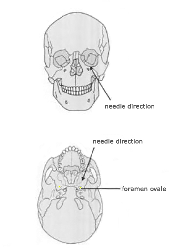
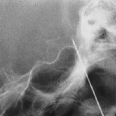
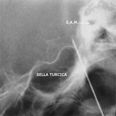
adapted from<a class="external" href="http://books.google.com.au/books?id=B4TY82NLR6sC&pg=PA687&lpg=PA687&dq=trigeminal+nerve+blocks&source=bl&ots=9Rxu-l2gzt&sig=kmzsxrFifzUJaHSMF_dI9MQ8-IU&hl=en&ei=Cb4pSuSKKJWMkAX1h7njCg&sa=X&oi=book_result&ct=result&resnum=12#PPA688,M1" rel="nofollow" target="_blank">Carol A. Warfield, Zahid H. Bajwa. Principles and Practice of Pain Medicine, 2004, p688</a>Lateral screening may be required to check the needle depth. The needle tip is likely to be correctly positioned when CSF is seen to leak from the needle. Contrast media is injected to show filling of the cistern.
When the needle tip position is confirmed to be correctly located, the patient is sat upright with the patient's head flexed to ensure that the injected glycerol remains within the infero-lateral portion of the cistern which contains the nerve rootlets of interest. Glycerol (a neuroleptic agent) is then injected. The needle is then withdrawn and the operator compares the patient's face for sensitivity bilaterally.
On completion of the procedure, the patient is transferred to his/her bed and seated upright (bed backrest up) for at least one hour of observation.
Technique Notes
- Check with the operator (radiologist/neurosurgeon) whether they require patient sedation for this procedure. This will need to be organised in advance.
- The anatomy of interest is very small. Use a small field size and cone the X-ray beam as tightly as practicable
- Keep the image intensifier as close as practicable to the patient to improve image quality and reduce radiation dose.
- A preliminary DSI image taken by the radiographer will demonstrate to the operator the anatomy of interest. In addition, the radiographer will be sure that the foramen ovale can be demonstrated prior to starting the procedure.
- Label the images to indicate which side was treated.
...back to the Wikiradiography home page
...back to the Applied Radiography home page
