The Abdominal Plain Film- Appendicitis
Jump to navigation
Jump to search
Introduction
Case 2
Case 3
Case 4
Summary
.........back to the applied radiography home page
Identifying an appendicolith can assist greatly in diagnosing appendicitis in patients presenting with acute abdominal pain. This page looks at the appendicolith and other signs of appendicitis.
Epidemiology
Aetiology
- Appendicitis is the most common infective condition that presents as an acute abdomen
Clinical Pathway
- Requires the occlusion of the appendiceal lumen by particulate material- material could be endogenously produced or foreign
- Material could be blocking the lumen or a focus or nidus upon which faeces adhere and calcium salts are laid down
Plain Film Signs of AppendicitisAn abdominal plain film is not considered an appropriate examination in patients with suspected appendiciis in many centres.
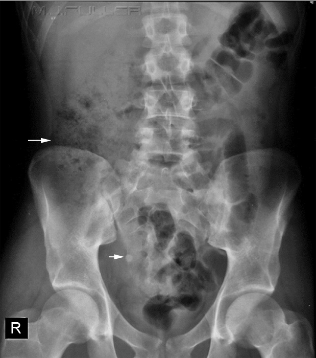
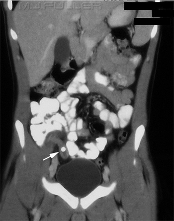
Case 1
Sign
Comment
Appendicolith
- Also known as coproliths, fecaliths and stercoliths
- Appendicoliths are common
- Can appear in various locations with variation in location of the appendix
- Not all appendix stones are calcified
Gas in Appendix
- This is not necessarily a sign of appendicitis- can be seen in the normal appendix
Abscess
RIF mass
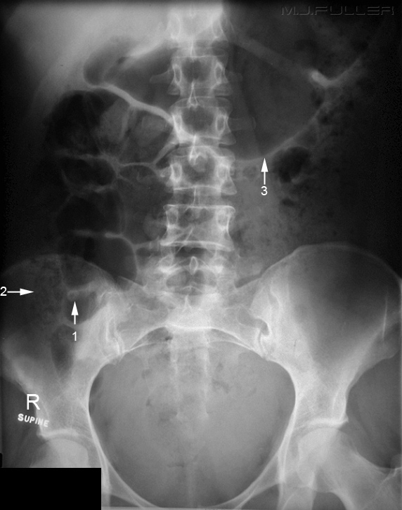
- There can be a general increase in opacity in the right lower quadrant associated with an appendiceal abscess
- Can also be seen as bubbles of gas in the abscess
Caecal Ileus
- Seen as dilated caecum
Flank Sign
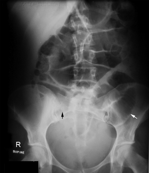
- Separation of the bowel from the right flank stripe by the lateral accumulation of pus and ascites
SBO
- Reflex ileus
Scoliosis
- Scoliosis of lumbar spine
•suggestion of a dilated caecum (top arrow)
Case 2
Case 3
Case 4
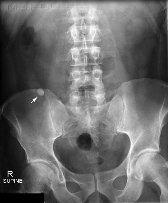
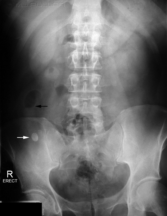
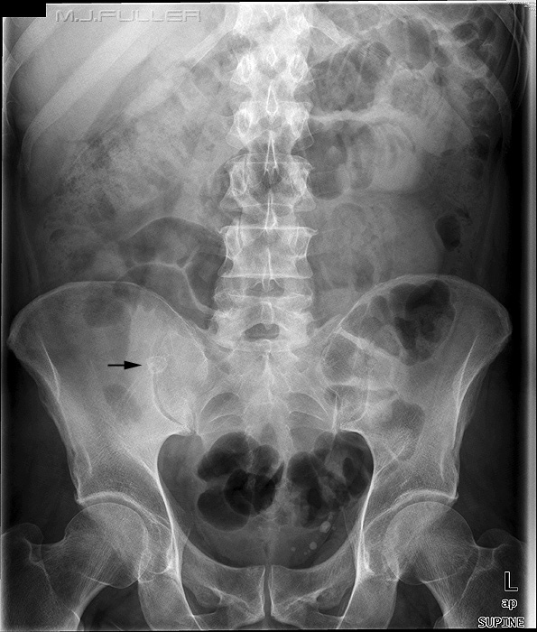
The arrowed structure could be mistaken for an appendicolith. It was revealed on CT to be a bony lesion in the pelvis. Not everything that looks like an appendicolith is an appendicolith!
Summary
An abdominal plain film diagnosis of appendicitis is not always possible. A significant proportion of appendicitis patients will have no plain film signs. However, when the signs of appendicitis are evident, a timely diagnosis will potentially improve patient outcomes. Importantly, the benefits of an abdominal plain film in patients with suspected appendicitis are questionable.
.........back to the applied radiography home page
