Sternum Radiography
Mechanisms of InjurySternum radiography is commonly performed in trauma centres. This page considers all aspects of sternal radiography and pathological appearances of sternal fractures.
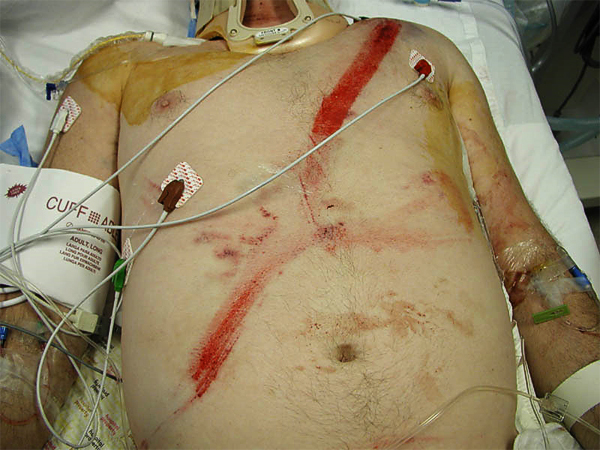
Seatbelt Injury
<a class="external" href="http://www.itim.nsw.gov.au/images/seat_belt_mark_2.jpg" rel="nofollow" target="_blank">http://www.itim.nsw.gov.au/images/seat_belt_mark_2.jpg</a>Whilst there are a myriad of possible mechanisms of injury for sternal fracture, seat belt injury deserves a special mention. In countries where the wearing of seatbelts in cars is compulsary for all occupants, this is a common sight for staff in the Emergency Department. The injury is caused by the rapid deceleration of the car during a motorvehicle accident, particularly in a frontal collison, where the occupants are forcibly propelled forwards onto the seatbelt. In these accidents, the occupants will present to the Emergency Department with a characteristic 'red sash' linear bruise or abrasion/petechiae that corresponds with the orientation of the seatbelt.
Lateral Sternum or Lateral Chest?
It is possible to produce a lateral sternum image by post-processing a digital lateral chest X-ray image. The results are variable in their effectiveness.
Case 1
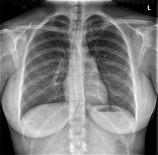
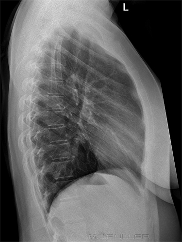
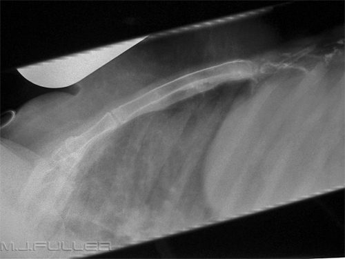
This patient presented to the Emergency Department following a motor vehicle accident. She was complaining of a painful sternum and was referred for radiography of the chest and sternum.
The radiographer considered the visualisation of the sternum on the lateral chest image to be inadequate for the lower sternum.
A dedicated erect lateral sternum was performed with a view to improving the visualisation of the lower sternum.
The dedicated lateral sternum image has achieved marginally improved visualisation of the lower sternum.
Using the Snake with the Lateral Sternum Projection
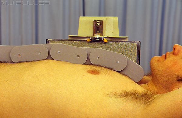
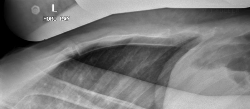
The snake was designed to be self-conforming to the chest wall when used on the horizontal ray lateral projection of the sternum as shown below.
Snake positioned for a Horizontal Ray Lateral Sternum Projection
The snake is easily kept upright using a blanket which is laid across the top of the patient and covering the lower segment of the snake (not shown)
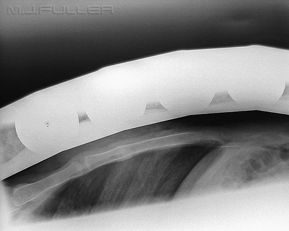
Lateral cross-table sternum using snake to reduce scatter radiationCase 1
This patient presented to the Emergency Department following a head-on motor vehicle accident. The patient was experiencing considerable sternal pain and was referred for radiography of the sternum. These images were taken using conventional film-screen radiographic equipment.
Image 1- No Snake
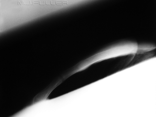
This is the initial lateral sternum image. It is apparent that the there is underexposure of the lower sternum due to overlying breast shadow. There is also overexposure of the manubrium. The radiographer considered that the image was degraded by scatter radiation. She kept the exposure factors the same and repeated the image using the snake positioned over the sternum
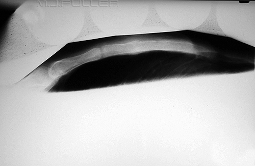
Image 2- Repeat View with Same Exposure with Snake In-situ
This repeat view of the lateral sternum is clearly of a much higher quality. The higher quality of this repeat image can be almost completely attributed to the reduction in scatter radiation associated with the use of the snake. A fracture of the sternum with possible associated retro-sternal soft tissue swelling is now revealed
Case 2
This patient also presented to the Emergency Department following a head-on motor vehicle accident. A trauma radiographic series was requested including lateral sternum. The radiographer thought that the patient's very large breasts would cause considerable artifact if a lateral cross-table sternum projection was attempted. The risk of failure was so high that conventional cross-table lateral sternum radiography was considered likely to fail.
Following discussion with the doctor in attendance, it was decided that a lateral cross table sternum projection would be attempted using the snake and with taping of the breasts so that they were positioned anterior to the sternum. The "taped up" breasts held the snake in position. The patient freely consented to the procedure.
The resulting lateral cross-table sternum image is shown. The results are remarkably good in a patient who appeared to be to be unsuitable for cross-table sternum radiography.
The distance between the visible snake segment and the sternum represents the thickness of the soft tissues anterior to the sternum. There is no evidence of breast tissue overlying the sternum.
Clearly, there must be explicit consent from the patient to undertake this procedure. The positioning of the snake and the taping of the breasts was undertaken by a female member of the radiography staff.
Case Studies
Case 1
Case 2
