Soft Tissue Signs- Thoracic Spine
The ability to recognise soft tissue signs in the chest can provide valuable clues to spinal and paraspinal pathology. This page focuses on the paraspinal line changes associated with spinal trauma.
Case Study 1
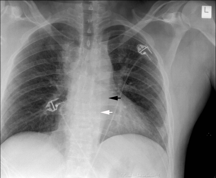
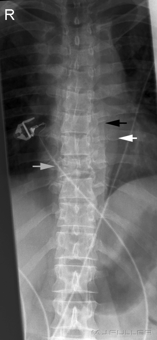
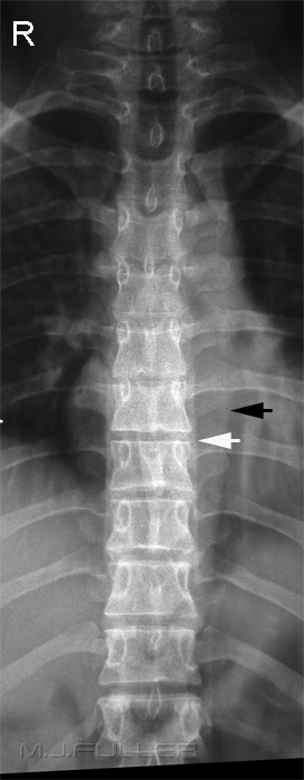
| | This patient presented to the Emergency Department following a motor vehicle accident. The black arrow identifies the descending aorta. The white arrow marks the left paraspinal stripe. "The left paraspinal line is formed by tangential contact of the left lung and pleura with the posterior mediastinal fat, left paraspinal muscles, and adjacent soft tissues. The left paraspinal line extends vertically from the aortic arch to the diaphragm and typically lies medial to the lateral wall of the descending thoracic aorta" (1) The left paraspinal line is normally in close proximity to the spine. This paraspinal line appears unusually wide and can indicate adjacent spinal pathology. Do the thoracic vertebral bodies appear normal? The patient cannot move- what additional imaging would you perform? |
Discussion
This case study illustrates the potential for a diligent radiographer to make a difference in the resus room. The abnormal paraspinal line was not identified by the doctor on duty in the resus room. The radiographer's successful identification of this soft tissue sign suggested a need for imaging of the thoracic spine. The successful and early identification of a spinal injury can change the course of the patient's management in those early minutes in the resus room. Following the identification of the T9 fracture the patient is assumed to have an unstable fracture until proven otherwise. The patient then becomes a "spinal patient" and all movements and transfers are made with this in mind.
References
(1) Jerry M. Gibbs, Chitra A. Chandrasekhar, Emma C. Ferguson, and Sandra A. A. Oldham
Lines and Stripes: Where Did They Go? —From Conventional Radiography to CT, RadioGraphics 2007;27:33-48
.... return to the Applied Radiography home page


