Soft Tissue Signs- The Wrist
Health professionals with expertise in plain film image interpretation will approach an equivocal fracture by critically examining the bony features in the context of other information such as
- Patient history
- Clinical presentation
- soft tissue signs
This page will examine the soft tissue signs of bony injury of the wrist- what they are, their plain film appearances, their limitations, and their utility.
Soft Tissue Signs
Curtis D.J. and Downey E.F. (Gilula, L.A. The Traumatised Hand and Wrist, Radiographic and Anatomic Correlation, 1992, p50) observed the folowing in relation to radiography of the wrist.
| Soft tissue swelling involving fat planes should conform to the area of underlying soft tissue or bone injury. "Swelling seen in an area not associated wih an observed fracture should initiate a search for an additional abnormality to explain the swelling. The possibility of a fracture or dislocation must be excluded with particular care, even though the routine views of the bones are normal in the presence of soft tissue swelling if more than one fat plane is affected or if the soft tissue swelling is unequivocal. The absence of soft tissue swelling virtually excludes the presence of a fracture, but the converse is not necessarily true... With definite swelling, additional views to better demonstrate the the area underlying the swelling should be considered" (Gilula, L.A. The Traumatised Hand and Wrist, Radiographic and Anatomic Correlation,, 1992, p50). | ||
Anatomy
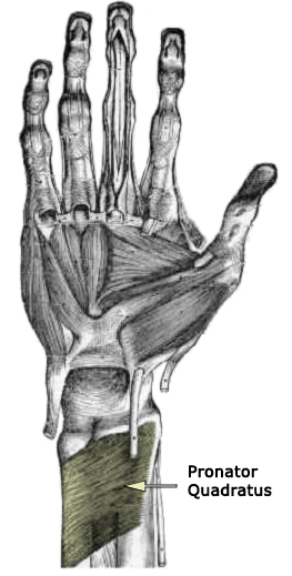
The Pronator Quadratus Muscle
adapted from from An Atlas of Human Anatomy by Carl Toldt, M.D., 1919."The pronator quadratus muscle is a small quadrilateral muscle attached to the anterior aspects of the distal sixths of the radius and ulna. A well-defined fascia covers the muscle, preventing intermuscular communication and creating a distinct forearm space in which fluid can accumulate (4). A thin layer of fat overlies the fascial covering, between the pronator quadratus and flexor digitorum profundus muscles (1,4). This fat plane can be seen as a thin radiolucency on at least 90% of lateral radiographs of normal distal forearms (1,3). The thin radiolucency can either have a slight anterior convexityor be straight and parallel to the ra-dius (1,3). In addition, the distal edge of the fat plane is in contact with the volar aspect of the radius (3). The pronator quadratus sign is visible when fluid, such as blood, accumulates within this muscle (1–4). If swollen, the pronator quadratus muscle bulges anteriorly and can displace or even efface the lucent fat plane (1–4)."
<a class="external" href="http://radiology.rsnajnls.org/cgi/reprint/244/3/927.pdf" rel="nofollow" target="_blank">
Josh B. Moosikasuwan,MD
The Pronator Quadratus Sign1
Radiology: Volume 244: Number 3—September 2007</a>
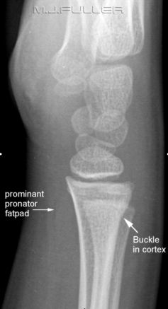
Pronator (Quadratus) Fat Pad Sign
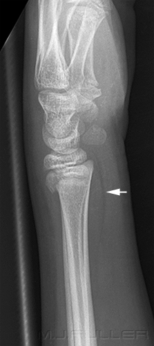
| | .- The dorsal cortex of the distal radius can be seen to deviate from a smooth curve suggesting a buckle fracture. Note that this is very subtle finding but was noted (and 'red dotted') by the radiographer at the time - A subtle lucent bony defect can be seen in the distal radius closely related to the buckled radial cortex - Subtle volar tilt or palmar inclination cannot easily be measured in this age group. There does not appear to be significant dorsal angulation of the radius -The pronator fat pad is raised suggesting a significant wrist injury (bulging of the fascial covering of the pronator quadratus muscle caused by haematoma) -A comparison view of the patient’s contralateral wrist is not required to arrive at a diagnosis |
.
Limitations of the Pronator Fat Pad Sign
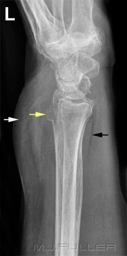
| | This is a lateral wrist image on an elderly patient who has presented to the Emergency Department following a fall onto her left wrist. There is a clearly visible fracture of the radius with cortical buckling on the dorsal aspect of the radius (yellow arrow). The pronator fat pad is clearly visible and appears to be normal. There is a very large soft tissue swelling seen over the dorsal spact of the wrist (white arrow). One study reported that in 50 cases of confirmed distal radius fractures, only 30 percent demonstrated a pronator fat pad sign. (1) |
Limitations of the Pronator Fat Pad Sign
References
1. G. Annamalai and N. Raby Scaphoid and Pronator Fat Stripes are Unreliable Soft Tissue Signs in the Detection of Radiographically Occult Fractures. Clinical Radiology. Volume 58, Issue 10, October 2003, Pages 798-800
Scaphoid Fat Pad Sign
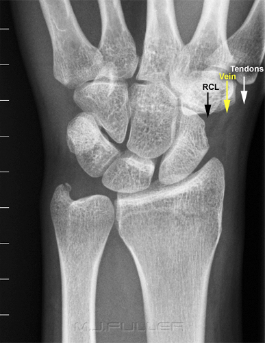
Scaphoid fractures are the most common fracture of the carpal bones, accounting for 70% of carpal bone fractures (1). Ninety percent of all acute scaphoid fractures heal if treated early. (1) The scaphoid fat pad sign refers to a propensity for distortion or obliteration of the juxtaposed fat stripes in the presence of a scaphoid fracture. The scaphoid fat pad is demonstrated best with the wrist in ulnar deviation.
| | 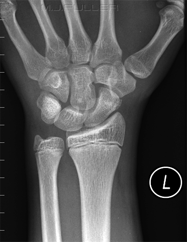 Obliteration of Scaphoid Fat Pad associated with fracture of the scaphoid tubercle |
A positive scaphoid fat pad sign does not definitely indicate an adjacent scaphoid fracture. In one study it was reported that scaphoid fracture was found on MRI scanning in only 50% of patients who demonstrated a positive scaphoid fat pad sign. (2)
.
References
... back to the Applied Radiography home page1. Carol A Boles, MD. Wrist, Scaphoid Fractures and Complications, Nov 16, 2007. <a class="external" href="http://www.emedicine.com/RADIO/topic747.htm" rel="nofollow" target="_blank">http://www.emedicine.com/RADIO/topic747.htm</a>
2. G. Annamalai and N. Raby Scaphoid and Pronator Fat Stripes are Unreliable Soft Tissue Signs in the Detection of Radiographically Occult Fractures. Clinical Radiology. Volume 58, Issue 10, October 2003, Pages 798-800
