Soft Tissue Signs- Knee Trauma
Introduction
An equivocal fracture should be considered in the context of other information such as
- Patient history
- Clinical presentation
- mechanism of injury
- soft tissue signs
This page will examine the soft tissue signs of bony injury of the knee- what they are, their plain film appearances, their limitations, and their utility.
The Knee
Soft Tissue Signs of Knee Effusion on Lateral View
- Suprapatellar pouch width greater than 5mm
- Blurring of posterior aspects of quadriceps tendon
- Increased soft tissue density (fluid density) around anterior femoral condyles (Hoffa's triangle)
- Widened joint space
- Bowing of Quadriceps tendon
- Anterior displacement of patella
- Bulging of Posterior fat lines
- Displacement of fabella
adapted from Weissman and Sledge, 1986, p522 (1)
Knee Effusion- Very Small
This patient presented with a history of a fall the previous day. There appears to be a small amount of fluid in the supra-patella pouch (left arrow). There is also fluid in Hoffa's Triangle (right arrow). Note that the knee is over-flexed. This position makes assessment of fluid in the supra-patella pouch more difficult.

Knee Effusion- Small
This patient presented with a history of dislocated right patella. There is a small knee effusion. There is a small amount of fluid in the supra-patella pouch (left arrow) and in Hoffa's Triangle (right arrow).

Knee Effusion- Moderately Large
The patient fell out of a tree onto both feet. There is a knee joint effusion. This is evident from the fluid density seen in the supra-patella pouch (left arrow) and in Hoffa's fat pad (right arrow). A knee effusion indicates an intra-capsular injury (but not necessarily a fracture). A knee effusion is not particularly specific or diagnostic, but the absence of a knee effusion can be useful in cases of equivocal fracture.

Knee Effusion- Very Large
This elderly patient fell onto her right knee. She has sustained a fractured patella and has a very large knee joint effusion (arrowed).

Knee Effusion- comparison
| Knee effusion  | Normal Knee |
| This patient has a knee effusion. Note the fluid in the suprapatellar pouch and in Hoffa's fat pad (black arrows). Note also the patella is displaced anteriorly and angled as a result of the pressure exerted by the fluid in the suprapatellar pouch. Compare this image with the patient's other knee-> | This is the contralateral knee on the same patient which shows no sign of knee effusion |
.
Estimating the size of knee Effusions
Hall (2) correlated the size of knee effusions on plain film with the amount of aspirated fluid.
Width of Base of Suprapatellar Pouch
Knee joint Aspiration Volume (ml)
<5 mm o 5- 10mm 1 to 9ml 10mm or greater 10ml or greater
Extra-Articular Swelling
This patient's history is unknown. There is evidence of soft tissue swelling on the medial aspect of the knee on the AP image (arrowed). There is also soft tissue swelling anteriorly on the lateral image. There is increased density demonstrated within Hoffa's Triangle. This probably represents overlying extra-articular swelling rather than a knee effusion. The supra-patella pouch is not distended by a knee effusion. The appearances are consistent with an extra-articular antero-medial soft tissue swelling.
Extra-articular Swelling
This elderly patient presented to the Emergency Department following a fall. He sustained injuries to his facial bones and his left knee was severely swollen. The knee swelling was largely localised over the patella and appeared to be all extra-articular. In particular, there was no obvious swelling of the suprapatellar pouch.
The skyline image was performed with the aid of a Leddra Cassette holder. There is an impressive soft tissue swelling largely localised over the patella. There is no evidence of knee joint effusion. Hoffa's fat pad appears normal. There is no evidence of fracture or dislocation.
The radiographer who performed this examination was not expecting to find a fracture or dislocation. While the clinical appearance does not exclude a fracture or dislocation, the soft tissue swelling appeared to be all extra-articular suggesting no joint-related bony injury. The swelling corresponds with the prepatellar bursa. The patient reported that the swelling was immediate after the injury suggesting that the prepatellar bursa has filled with blood following the trauma.
The Knee Effusion Trap


| | |
|

Lipohaemarthrosis
A lipohaemarthrosis refers to the presence of a blood and fat in a joint (i.e. lipo= fat, haemo = blood, throsis= pertaining to a joint). The lateral horizontal ray knee view is the one knee projection where we see this appearance. A knee lipohaemarthrosis indicates that there is a fracture that communicates with the knee joint. The blood and fat are both associated with the fracture. The fat has entered the joint from the bone medulla via the fracture. The fat 'floats' on the blood resulting in a visible interface (red arrow).
It has been suggested that a patella fracture cannot cause a lipohaemarthrosis because there is insufficient fat within the patella. MR imaging of the patella (fat sat) demonstrates fat in the patella. I have seen lipohaemarthrosis associated with fractured patella on many occasions and not demonstrated any other bony injury. I am increasingly convinced that a fractured patella can result in lipohaemarthrosis of the knee which is visible on the horizontal ray lateral image.

<a class="external" href="http://www.gentili.net/FBI/knee_-_lateral_radiograph.htm" rel="nofollow" target="_blank">http://www.gentili.net/FBI/knee_-_lateral_radiograph.htm</a>This is an erect lateral knee. A lipohaemarthrosis is demonstrated. This is a potentially useful view for subtle lipohaemarthrosis because there is no superimposed quadriceps tendon. Its usefulness is limited by the likelihood that a patient with a knee fracture will not mobilise readily.

Lipohaemarthrosis Case Study
This patient presented to the Emergency Department following a sports injury the previous day. The patient sustained a blow to the knee and is now UTWB and is in great pain. The initial AP and lateral knee images are shown below
The Radiographer has performed AP and lateral horizontal ray knee views as shown above. There is a suggestion of a bony defect in the tibia overlying the fibula head (not marked). There is also evidence of a lipohaemarthrosis on the lateral image (white arrow). The fat/blood interface is not seen sharply. The radiographer decided to repeat the lateral view to try and confirm the presence of a lipohaemarthrosis.
The lipohaemarthrosis is now clearly seen. (the reason for the improved visualisation of the fat/blood interface on this repeat view is unclear)
The confirmation of the lipohaemarthrosis indicates that the patient does have a fracture that communicates with the knee joint. Given that the site and nature of the fracture had not been firmly demonstrated, the radiographer proceeded to perform oblique views of the knee.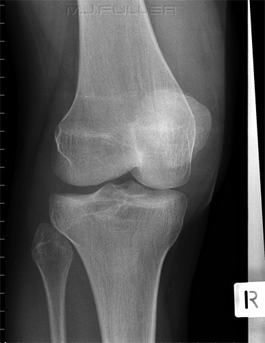


The external oblique image demonstrates a tibial plateau fracture of the lateral tibial condyle (white arrow).
Discussion
This case study demonstrates a clinical rather than a photographic approach by the radiographer. The x-ray examination was a process rather than an event. The images were taken in three stages with each stage building on the findings from the previous image(s).This is a more satisfying and effective approach than uncritically performing the routine views without further consideration of the findings.
? Loculated Lipohaemarthrosis
There is a lipohaemarthrosis of the knee joint. There appears to be three fat-blood levels in a stepped pattern (white arrows). This is occasionally seen and is possibly due to loculation of the suprapatellar space.

Case StudyRuptured Quadriceps Femoris Tendon
The quadriceps femoris tendon attaches the quadriceps muscle to the patella. This patient has had an unknown injury to the knee causing rupture of the quadriceps femoris tendon.
Note the unusual low position and forward tilting of the patella. The quadriceps tendon which is usually visualised contrasted by the suprapatellar fat body is not demonstrated(arrow) Quadriceps tendon re-attached post surgery. see also http://www.wikiradiography.com/page/Quadriceps+Tendon+Rupture
Ruptured Patellar Tendon
The patellar tendon attaches the patella to the upper tibia at the tibial tuberosity .
You will need to use your imagination with this one. You can see that the effect of complete rupture of the patella tendon is the opposite of that seen with the quadriceps tendon rupture. The patella is pulled superiorly by the quadriceps muscle<a class="external" href="http://radiographics.rsnajnls.org/cgi/reprint/14/6/1191?maxtoshow=&HITS=80&hits=80&RESULTFORMAT=&fulltext=knee+ligament&searchid=1&FIRSTINDEX=0&sortspec=relevance&resourcetype=HWCIT" rel="nofollow" target="_blank">Gerald WCapps, MD, Curtis WHayes, MD. Easily Missed Injuries
around the Knee. RadloGraphics 1994; 14:1191-1210</a>
Torn Patellar Tendon
An incomplete tear will not cause the patella to retract superiorly as shown above.
This 48 year old lady fell onto her left knee and sustained a deep laceration below her knee. The patellar tendon appears to have sustained a significant tear. It is unlikely that the tendon has completely ruptured given that it has remained in a normal alignment/position.
Note that there is air in the joint (Hoffa's fat pad) and there also appears to be small fragments of dirt associated with the laceration.<a class="external" href="http://radiographics.rsnajnls.org/cgi/reprint/14/6/1191?maxtoshow=&HITS=80&hits=80&RESULTFORMAT=&fulltext=knee+ligament&searchid=1&FIRSTINDEX=0&sortspec=relevance&resourcetype=HWCIT" rel="nofollow" target="_blank">
</a>


This case study attempts to demonstrate how a knowledge of soft tissue signs can help with identification and demonstration of a fracture.
This patient has presented to the Emergency Department after a fall onto her left knee. The patient has been referred for a knee X-ray examination. The patient's patella is tender but there is very little soft tissue swelling.
There appears to be a vertically orientated lucency through the medial aspect of the patella. This could represent a new fracture, an old fracture, or a bipartite patella.
This is an uncommon site for a bipartite patella (more commonly sited supero-lateral).
There is possibly a small joint effusion (see Hoffa's fat pad and the suprapatellar pouch)
There is no obvious soft tissue swelling overlying the patella but the image is a little overexposed for the soft tissues (pre-digital image)
There is evidence of a corticated margin to the "fracture" suggesting that it is not an acute injury.


Discussion
The AP view image demonstrates a possible patella fracture. The two persuasive detractors are
- the patient does not have a fractured patella clinically
- the images do not demonstrate a significant knee effusion or other soft tissue swelling that you would expect with this type of fracture.
Conclusion- bipartite patella or old ununited fracture. Note that a bipartite patella is more commonly seen on the lateral aspect of the patella.
Links
<a class="external" href="http://www.cs.sfu.ca/%7Ehamarneh/ecopy/spiemi2005b.pdf" rel="nofollow" target="_blank">Ghassan Hamarneha¤, Vincent Chua, Marcelo Bordalo-Rodriguesb, Mark Schweitzerc </a><a class="external" href="http://www.cs.sfu.ca/%7Ehamarneh/ecopy/spiemi2005b.pdf" rel="nofollow" target="_blank">Deformation Analysis of Hoffa’s Fat Pad from CT images of Knee Flexion and Extension</a>
http://www.wikiradiography.com/page/Quadriceps+Tendon+Rupture
References
1. Weissman, B and Sledge, C. Orthopedic Radiology. Saunders and Company, 1986
2. Hall FM. Radiographic diagnosis and accuracy in knee joint effusions.
Radiology 1975;1 15:49-54
.... back to the radiography home page here