Difference between revisions of "Soft Tissue Signs- Cervical Spine"
(Created page with "<div class="WPC-editableContent"><font size="4"><b>Introduction</b></font><br/><blockquote> Cervical spine imaging is one of the traditional mainstays of trauma radiography. T...") |
(No difference)
|
Latest revision as of 17:41, 11 November 2020
Cervical spine imaging is one of the traditional mainstays of trauma radiography. The lateral cervical spine view is usually a routine view in patients with severe trauma (although increasingly displaced by CT imaging). The emphasis on 'clearing' the cervical spine in major trauma patients reflects the importance of not missing cervical spine injuries. This page considers soft tissue signs of cervical spine trauma.
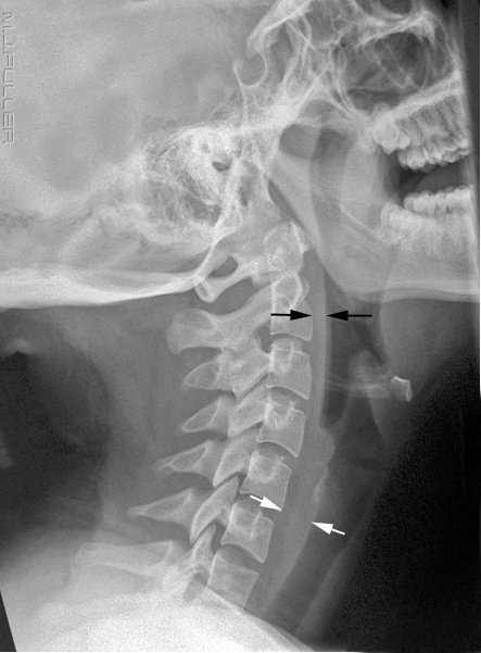
| | The retropharyngeal/preveretebral soft tissues can provide signs of cervical spine injury. This image demonstrates normal preveretebral soft tissues Two assessments of prevertebral cervical spine soft tissues are commonly made. These criteria are guides only. Various studies have suggested alternative guidelines and any abnormal soft tissue findings should be interpreted in the context of bony appearances, mechanism of injury and clinical signs. |
Pseudomass 1
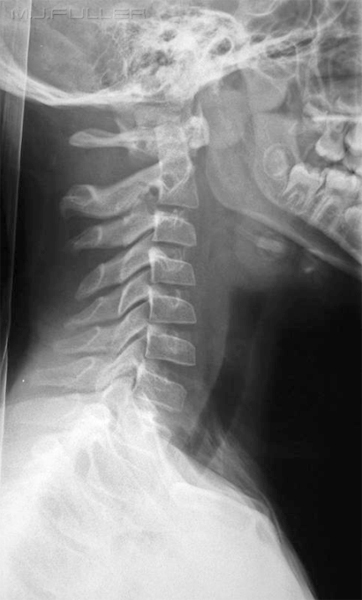
The assessment of the prevertebral cervical soft tissues can be impossible if the patient (particularly paediatric patients) is swallowing at the time of exposure.
Pseudomass 2
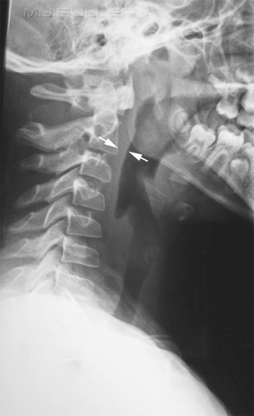
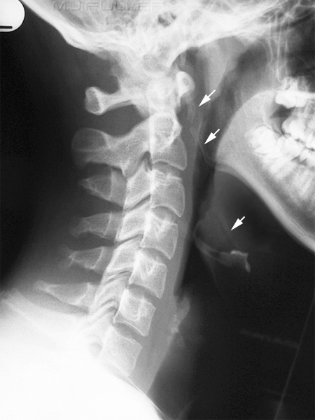
| | This is an adult patient who was probably imaged mid-swallow. The prevertebral soft tissues associated with C1 and C2 appear abnormally wide. This appearance is associated with the patient swallowing rather than any cervical injury. Radiographers use various techniques to avoid this appearance including exposing on inspiration and asking the patient to breathing through an open mouth at the time of exposure (try swallowing while breathing through an open mouth). Alternatively, asking the patient not to swallow at the time of exposure can be effective. |
The Cervical Pseudo-bone Tumour
Stylohyoid Ligament Calcification
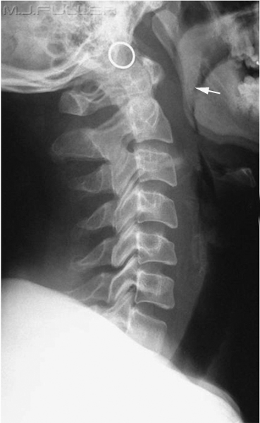
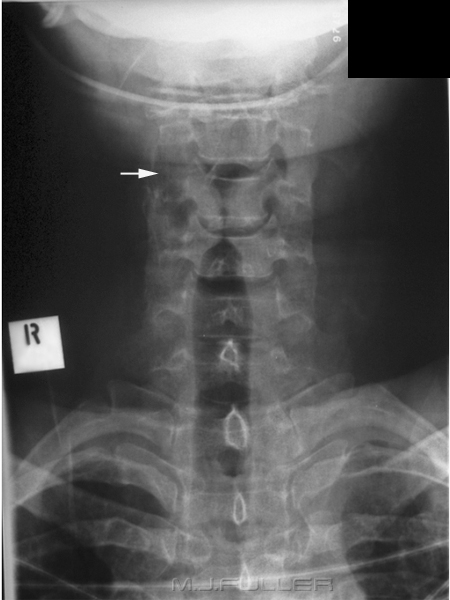
| | The arrowed bony structure is calcification of the stylohyoid ligament. In florid cases this calcification can involve the entire stylohyoid ligament bilaterally. Extensive calcification of the stylohyoid ligament is associated with Eagle Syndrome. |
References
1. Susan D. John, MD and LeonardE. Swiscbuk, MD Stridor and Upper Airway Obstruction in Infants and Children.RadioGraphics, 643, July 1992
...back to the Applied Radiography home page
