Salter-Harris Fractures
</a>
Introduction
It is important for radiographers to be able to identify fractures that involve the growth plate. These fractures can have significant implications for treatment and prognosis. Furthermore, identification of the type of growth plate trauma can have implications for treatment and prognosis. This page examines the Salter-Harris classification of fractures involving the growth plate and the radiographic techniques for demonstrating them
The Salter-Harris Classification

Source: <a class="external" href="http://www.primary-surgery.org/ps/vol2/html/sect0324.html" rel="nofollow" target="_blank">Epiphyseal injuries in children</a>
The Salter-Harris fractures are classified 1 - 5. The higher the classification number, the worse the prognosis. Most Salter Harris injuries do not result in bone growth disturbance. If the epiphyseal plate is injured, the volar portion of the epiphyseal plate closes before the dorsal portion. (Kraemer B.A. and Gilula, L.A., The Traumatised Hand and Wrist, Radiographic and Anatomic Correlation,, 1992, pp165-166).
Long Bone Anatomy
- Diaphysis
- Metaphysis
- physis or epiphyseal plate
- epiphysis
Elbow
Lateral Condyle Fractures
Salter notes that the lateral condyle fractures of the distal humerus are Salter Harris 4 fractures..
<a class="external" href="http://books.google.com.au/books?id=oa6fDFuX-I8C&pg=PA523&lpg=PA523&dq=elbow+condyle+vs+epicondyle&source=bl&ots=lhiHITotg0&sig=YDhHTE7q_t_nQN0pcYGHEr0kFqo&hl=en&ei=wSBPSrS3Loj8sQPOvYWrDQ&sa=X&oi=book_result&ct=result&resnum=7" rel="nofollow" target="_blank">(Robert. Bruce. Salter ,</a>
<a class="external" href="http://books.google.com.au/books?id=oa6fDFuX-I8C&pg=PA523&lpg=PA523&dq=elbow+condyle+vs+epicondyle&source=bl&ots=lhiHITotg0&sig=YDhHTE7q_t_nQN0pcYGHEr0kFqo&hl=en&ei=wSBPSrS3Loj8sQPOvYWrDQ&sa=X&oi=book_result&ct=result&resnum=7" rel="nofollow" target="_blank">Textbook. of Disorders and Injuries of the Musculoskeletal System. 3rd Ed, 1999, p523)</a>

This is an attempted AP elbow image that has been over-rolled into an oblique position. The image is under-exposed resulting in a low signal-to-noise ratio. There is a fracture of the lateral condyle of the distal humerus. This fracture involves the epiphysis, epiphyseal plate and lateral metaphysis/condyle. This is a Salter Harris IV fracture.
The integrity of the reminder of the epiphyseal plate is unclear.

Table of Salter Harris Fractures
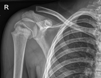
Anatomy SH I SH II SH III SH IV SH V Proximal Humerus
Salter-Harris I 'Fractures' of the Distal Fibula
This 7 year old boy presented to the Emergency Department following a fall from play equipment. He was unable to weight-bear (UTWB) and his right ankle was painful and swollen.
There is considerable swelling over his distal fibula.The anterior and posterior recesses of the ankle joint (arrowed) are of fluid density and suggest the presence of a large ankle effusion. The age-group, the soft tissue swelling, the inability to weight-bear and the ankle effusion suggest a significant force and raise the possibility of a Salter Harris I injury to the distal fibular growth plate.
A Salter Harris I injury to the distal fibular epiphyseal plate will often not be definitively diagnosed on plain film. The diagnosis should be made on a balanced view of the clinical and radiographic evidence. Point tenderness over the distal fibular growth plate is an important diagnostic clue.

