Radiography of Subtle Wrist Fractures
Wrist fractures are occasionally very difficult to demonstrate radiographically, indeed, they are sometimes evident on MRI scanning or bone scan only. This page looks at some cases of tricky wrist fractures and the techniques for demonstrating them with plain film radiography. As is often the case, a clinical approach to radiography will yield better results than a photographic approach.
Case 1a
Case 1b
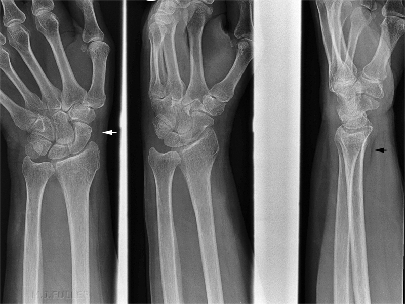
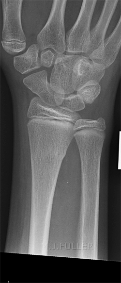
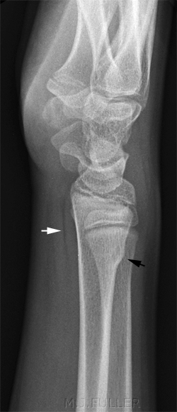
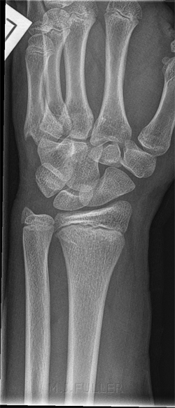
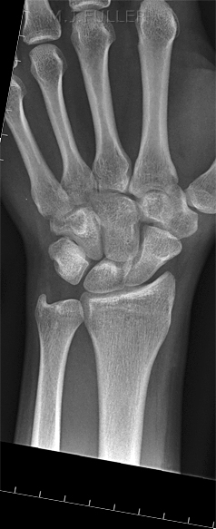
Routine wrist views were performed
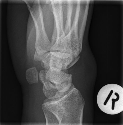
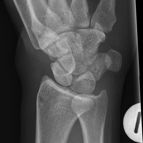
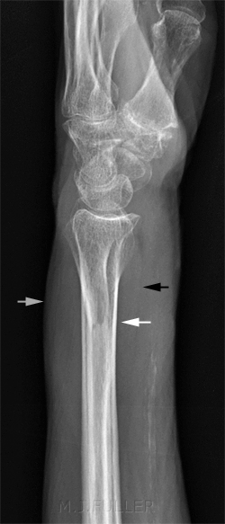
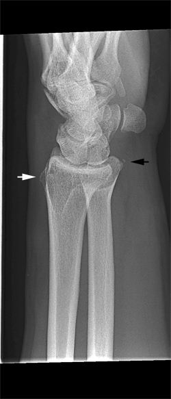
This series demonstrated no obvious fracture. Moreover, the scaphoid fat pad (white arrow) and the pronator fat pad (black arrow) were normal.
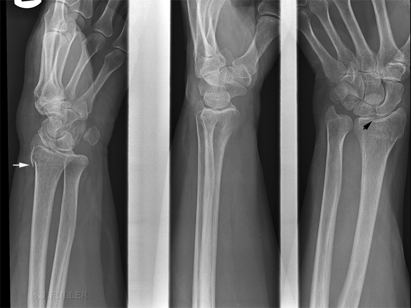
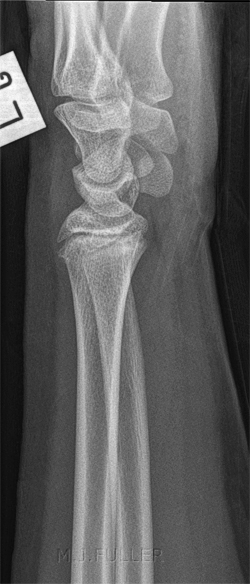
The radiographer was convinced clinically that the patient had sustained a fracture and considered whether the soft tissue signs were misleading (as there are known to be on occasions). Supplementary views were performed as shown below.
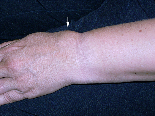
The 40 year old lady presented to the Emergency Department after falling over her dog. She was examined and was found to have a very painful right wrist. In addition, there was localised swelling over the dorsum of her wrist. She was referred for right wrist radiography.
Case 2
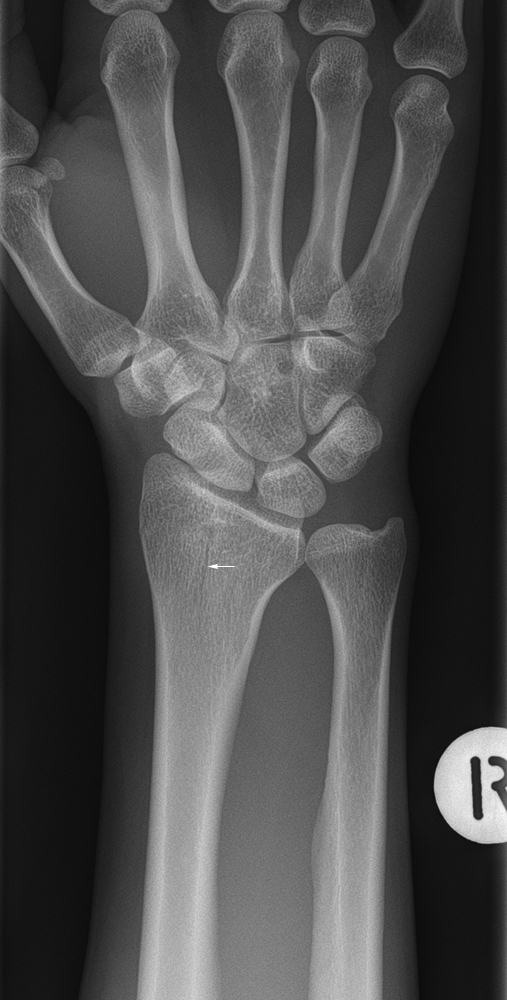
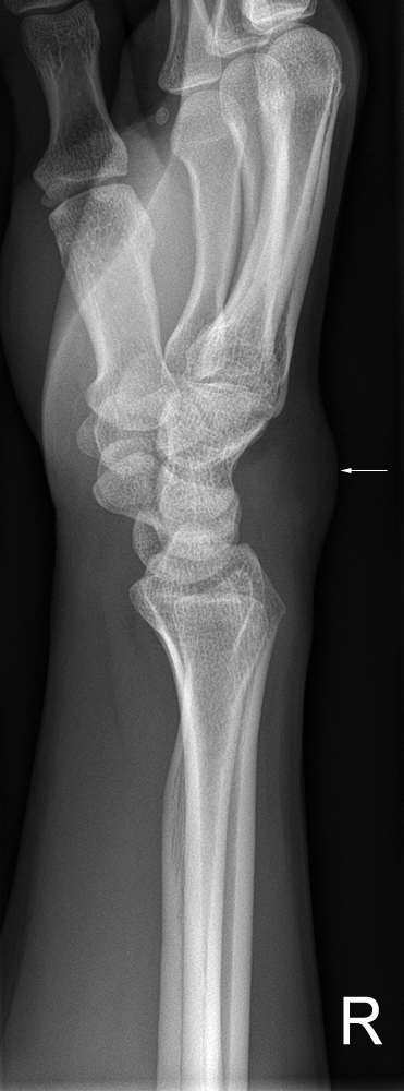
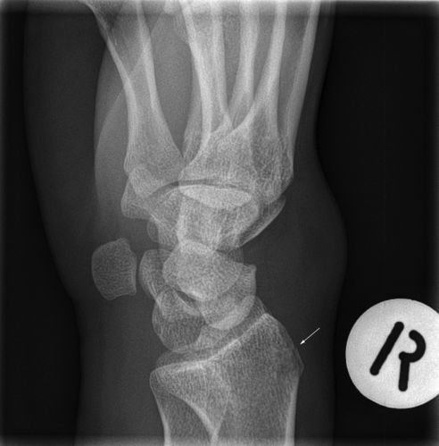
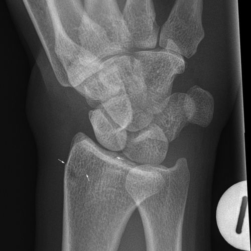
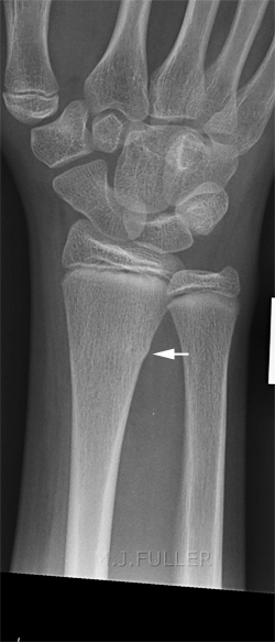
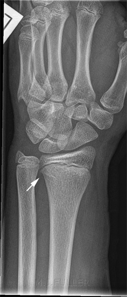
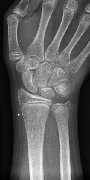
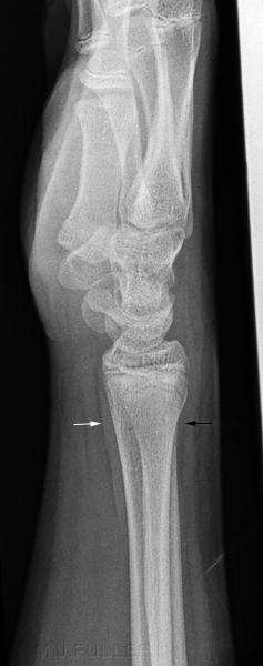
There is an impression of a vertical lucent line through the distal radial metaphysis. There is an impression of a vertical lucent line through the distal radial metaphysis (arrowed).
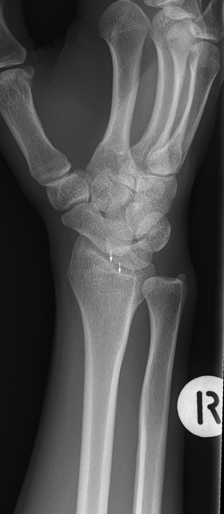
There appears to be a step in the articular surface of the distal radius. There appears to be a step in the articular surface of the distal radius (arrowed)
There is evidence of localised swelling over the dorsal aspect of the wrist There is evidence of localised swelling over the dorsal aspect of the wrist (arrowed)
IntermissionThe radiographer thought that the patient had sustained a wrist fracture based on clinical evidence (pain, swelling, limitations of movement). The radiographic evidence of wrist fracture on the routine projection images was inconclusive- on balance, supplementary projections were warranted
CommentThis is a good example of trauma radiography. The radiographers clinical assessment was the key factor that led to demonstration of the fracture. The radiographer was not satisfied that the routine projections had demonstrated a fracture conclusively in a patient who had convincing clinical signs of fracture.
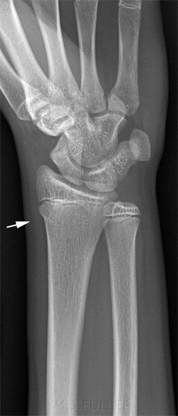
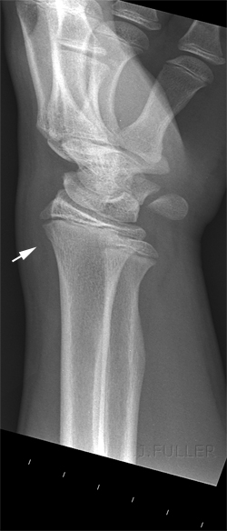
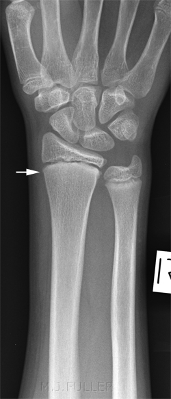
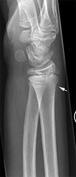
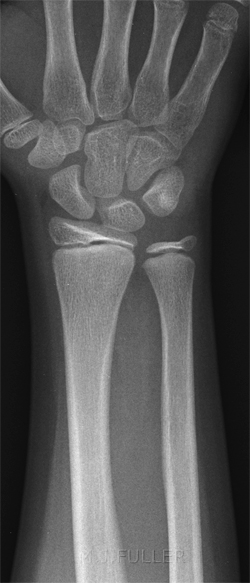
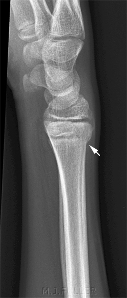
This 14 year old boy presented to the Emergency Department after a fall from his skateboard and was referred for wrist radiography. On examination, he had a painful wrist.
Comment
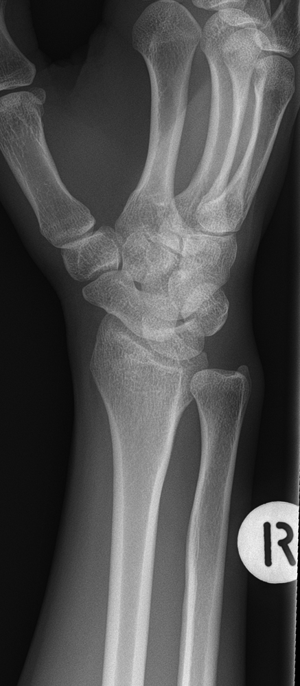
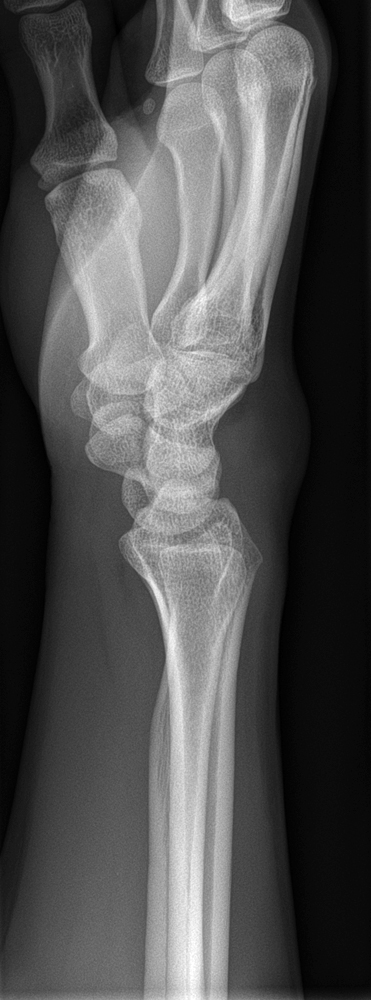
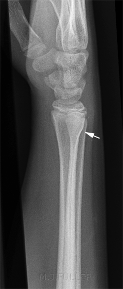
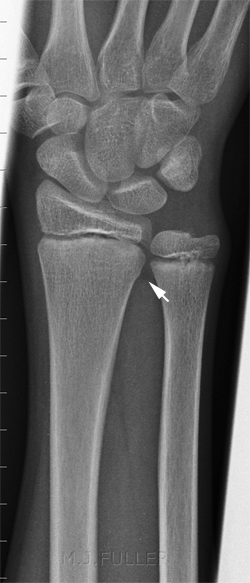
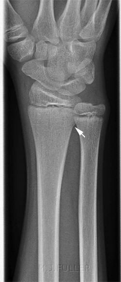
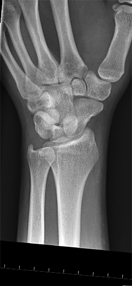
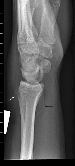
This is a very subtle fracture of the distal radius. There is a suggestion of dorsal palmar tilt on the lateral view although this is not convincing (zero degrees of palmar tilt can be a normal finding). Irregularity of the distal metaphysis immediately adjacent to the physis is normal. However, this does not appear to be typical of this commonly seen normal feature. As is often the case, the non-routine view (reverse oblique) demonstrates the cortical buckle most convincingly.
Case 3
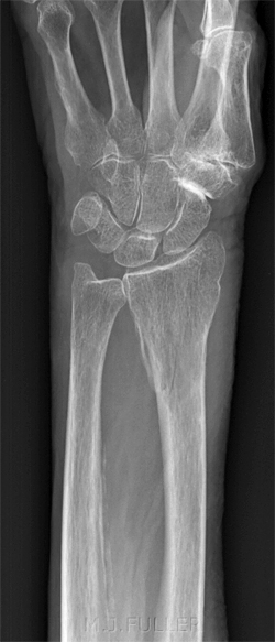
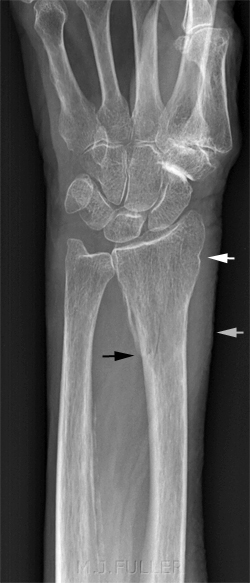
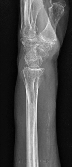
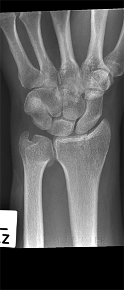
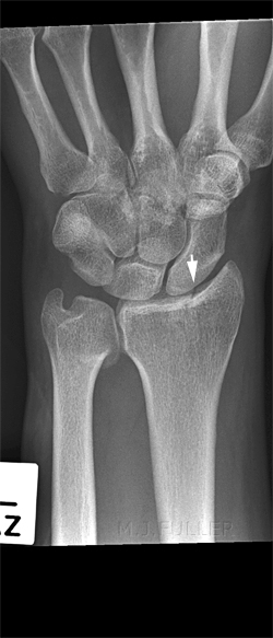
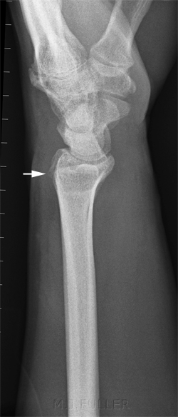
This 87 year old lady presented to the Emergency Department after a fall onto an outstretched hand. On examination she had a painful swollen wrist.
Comment
Case 4
Case 5This14 year old boy presented to the Emergency Department after a fall onto an outstretched hand. On examination he had a painful swollen wrist.
Comment
This14 year old girl presented to the Emergency Department after a fall onto an outstretched hand while playing netball. On examination she had a painful wrist.
Case 6Comment
This16 year old boy presented to the Emergency Department after a fall onto an outstretched hand. On examination he had a painful wrist.
Comment
Case 7
This14 year old boy presented to the Emergency Department after a fall from his pushbike. On examination he had a painful swollen wrist.
Comment
Case 8
This13 year old boy presented to the Emergency Department after a fall onto an outstretched hand.
Case 9Comment
This 17 year old boy presented to the Emergency Department after a fall onto an outstretched hand.
Comment
Case 10
This 45 year old man presented to the Emergency Department after a fall onto an outstretched hand.
Case 11
This 31 year old man presented to the Emergency Department after a fall onto an outstretched hand.
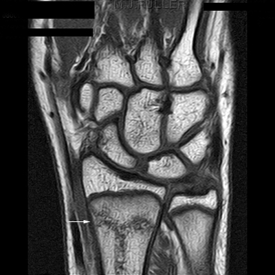
<embed allowfullscreen="true" height="350" src="http://widget.wetpaintserv.us/wiki/wikiradiography/widget/youtubevideo/907c53b0de96f656eb00ac0d8b3c92b88bd3558c" type="application/x-shockwave-flash" width="425" wmode="transparent"/> No convincing evidence of a fracture There is some loss of smooth contour of the radial aspect of the radial styloid. This is not convincing of a fracture There is a loss of continuity of the dorsal aspect of the radial metaphysis. There also appears to be a pronator quadratus soft tissue sign. The radiographic findings were considered to be equivocal. The patient's symptoms persisted. The referring doctor considered that the patient may have sustained a fractured scaphoid. The patient was referred for MRI scanning which revealed a radial metaphysis transverse fracture.
Case 12
This 13 year old boy presented to the Emergency Department after a fall onto an outstretched hand while running backwards.
Case 13
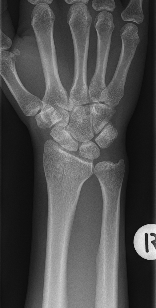
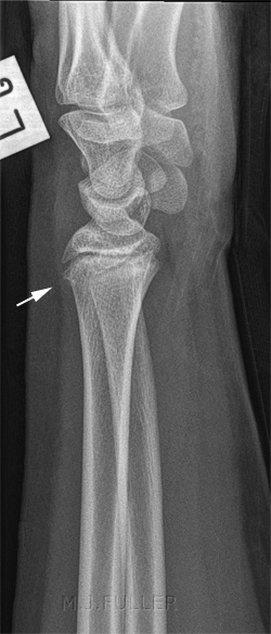
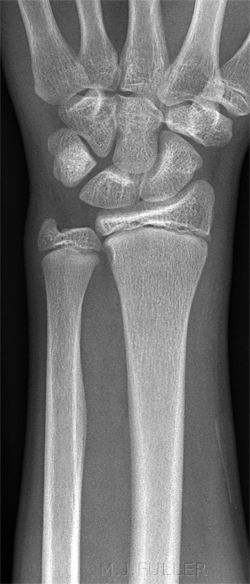
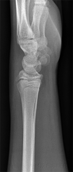
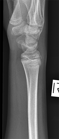
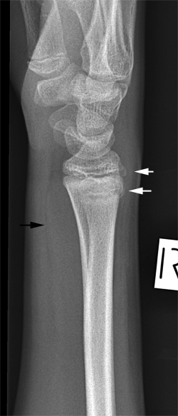
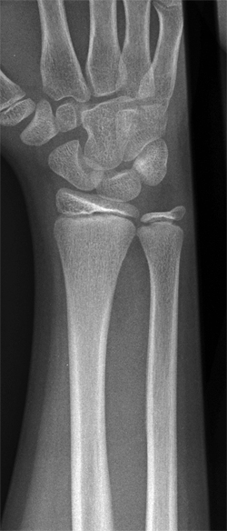
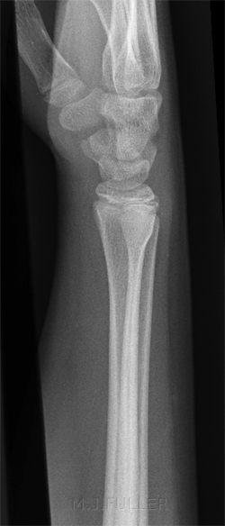
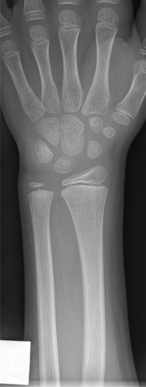
This 10 year old boy presented to the Emergency Department after a fall from his pushbike and was referred for wrist radiography. On examination, he had a painful wrist.
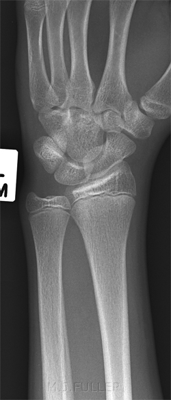
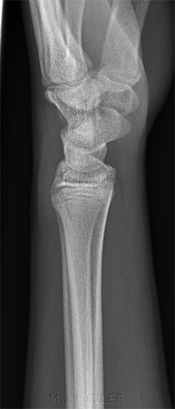
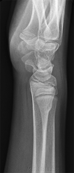
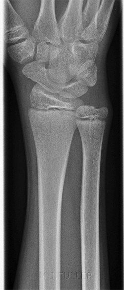
The PA wrist shows no evidence of displaced fracture There oblique wrist projection image shows no evidence of displaced fracture The lateral wrist projection image is not clearly abnormal.
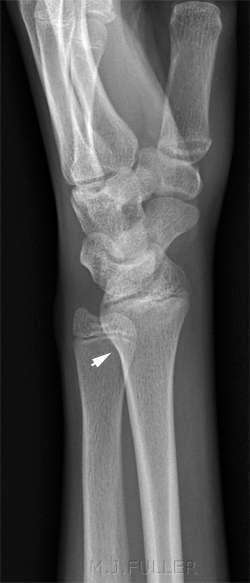
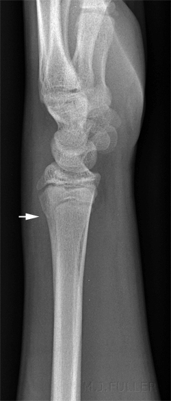
CommentThe radiographer noted the dorsal angular displacement of the distal radius and considered the possibility of a subtle distal radius fracture. A repeat off-lateral wrist projection was considered justified.
The off-lateral wrist projection image demonstrates a buckle fracture of the dorsal aspect of the radial metaphysis. Buckle fracture arrowed.
Comment
The radiographer should have a high level of willingness to undertake what would normally be considered poor quality radiography in order to demonstrate a suspected pathology. The off-lateral projection is a case-in-point.... doctrinaire thinking can be at the detriment of the diagnosis.
Discussion
The radial and ulnar cortical contours should appear as smoothly curved lines on plain film images of the wrist. . Any appearance other than a smooth curve should be considered as a possible fracture. The radial metaphysis in children and adolescents is fertile territory for subtle buckle fractures. Supplementary views such as reverse oblique and off-lateral can be useful in confirming the presence of a fracture. A clinical approach by the radiographer is particularly important- the radiographer should consider the patient's mechanism of injury, the clinical presentation and the soft tissue signs (clinical and radiographic).
Is the diagnosis of these fractures important? The simple answer is yes. A patient who has sustained a subtle buckle fracture will be treated with an immobilising plaster which will protect the fractured bone from further injury and reduce pain. Importantly, the patient's pain will be accounted for and appropriate pain relief prescribed. The patient will be assured that their symptoms have been explained and the patient be unlikely to represent for further diagnosis/treatment. The fracture can be followed up at a subsequent orthopaedic clinic appointment to ensure that it was not more serious than originally thought and remains minimally displaced. Appropriate limitations of activity can be explained to the patient to reduce the possibility of re-injury while the bone is healing. I also believe that there is an innate benefit in getting the diagnosis correct- satisfying for the patient, satisfying for the radiographer, and reassuring for the referring clinician. A more subtle benefit to the radiographer will be in the form of admiration and trust from the referring doctor- the radiographers can be left to their own devices if their radiography and judgements can be relied upon.
...back to the Applied Radiography home page
