Radiography of Pubic Rami Fractures

From wikiRadiography
Revision as of 17:34, 11 November 2020 by Travis (talk | contribs) (Created page with "<div class="WPC-editableContent"><font size="5"><b>Introduction</b></font> <br/><blockquote> Patients referred for hip radiography with suspected neck of femur fracture can s...")
(diff) ← Older revision | Latest revision (diff) | Newer revision → (diff)
Jump to navigation Jump to search
Introduction

Patients referred for hip radiography with suspected neck of femur fracture can sometimes by suffering from pubic rami fractures rather than neck of femur (NOF) fracture. This page considers radiographic techniques and pathological appearances of pubic rami fractures.

Radiography
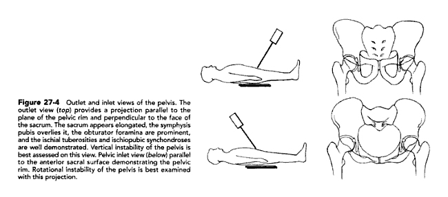
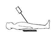
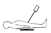
inlet and outlet views
Source:John J. Callaghan, Aaron G. Rosenberg, Harry E. RubashThe adult hip, Volume 1http://books.google.com.au/books?id=CSaFS5Tod3QC&pg=PA360&dq=lateral+hip+radiography&cd=9#v=onepage&q=lateral%20hip%20radiography&f=false
An easy memory aid to distinguish between the caudal angle and cephalic angle projections is to remember that the cephalic angle projection projects the rami towards the sacrum.


Case 1

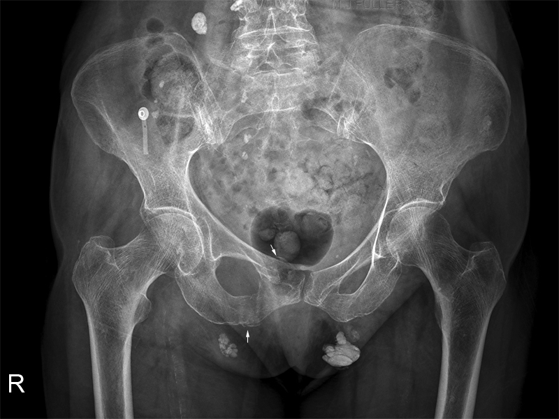
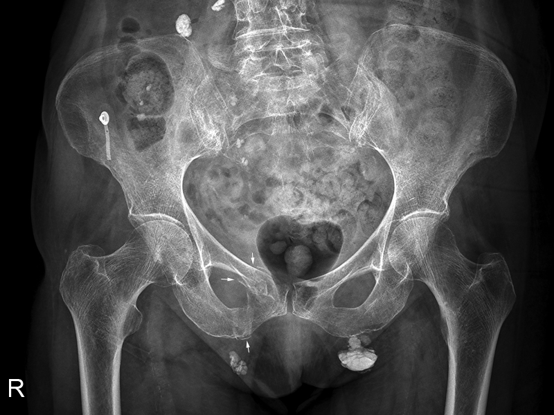
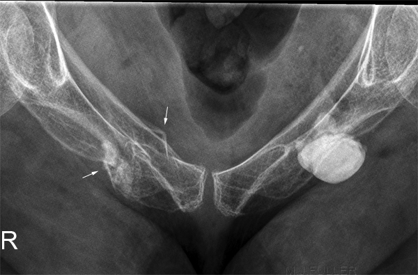
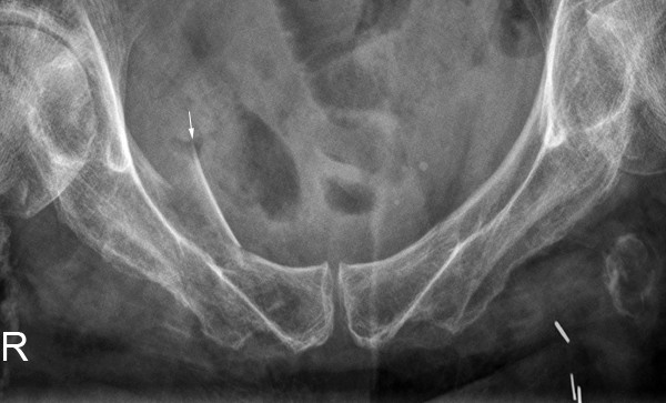
ap pelvis This 85 year old lady Presented to the Emergency Department after falling over when alighting from a bus. She was experiencing significant pain in her pelvis and was referred for radiography of her pelvis.

The initial AP pelvis image is shown left. The pelvis is rotated LOP. There was evidence of right sided pubic rami fractures. The radiographer considered a repeat view worthwhile with correction of the pelvic rotation.
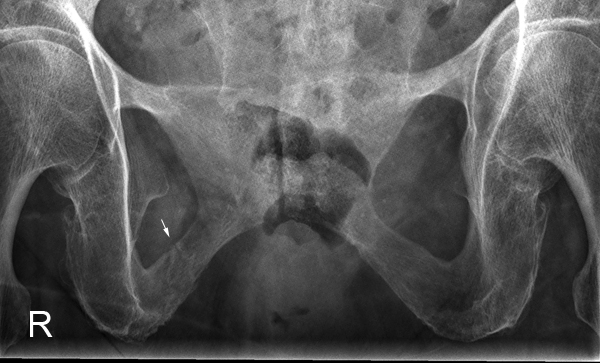
AP pelvis The right sided pubic rami fractures showed improved visualisation on the repeat AP pelvis image.

The calcific densities are of unknown aetiology but may be sequelae of previous lymphangiography.
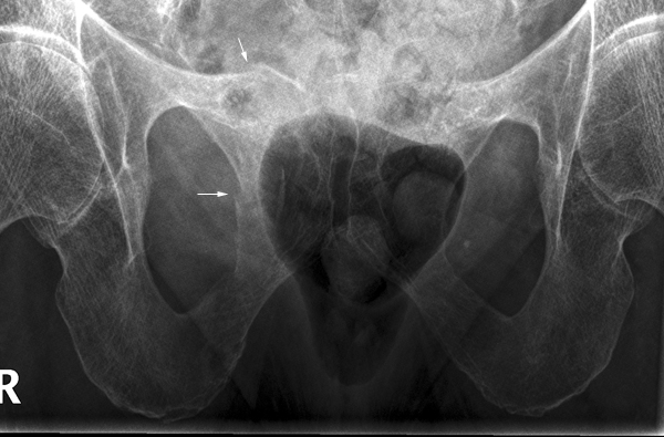
pubic ramiRadiography of Pubic Rami Fractures - wikiRadiography As is often the case, the caudal angulation pubic rami image best demonstrates the pubic rami fractures (arrowed)
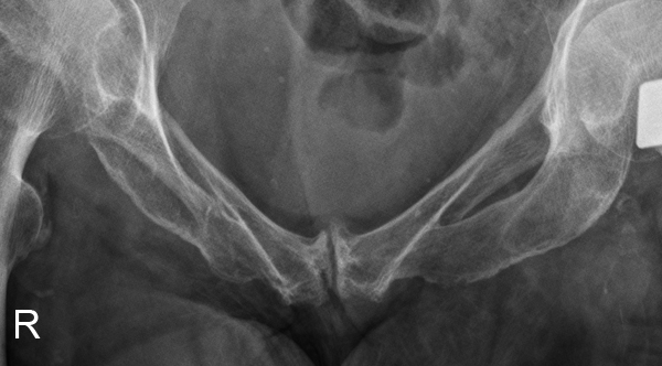
pubic ramiRadiography of Pubic Rami Fractures - wikiRadiography The cephalic angulation pubic rami view image demonstrates the right sided pubic rami fracture


Case 2
AP plevisThis 90 year old female presented to the Emergency Department after a fall. She was examined and subsequently referred to the Emergency Department with a provisional diagnosis of right neck of femur fracture.

The patient's pelvis is not in a true AP position (pelvis is slightly RPO). There is a suggestion of a minimally displaced inferior pubic rami fracture on the right (arrowed). Care must be taken with diagnosis of fractures in this region because of the common appearance of a normal appearance mimicking a fracture.
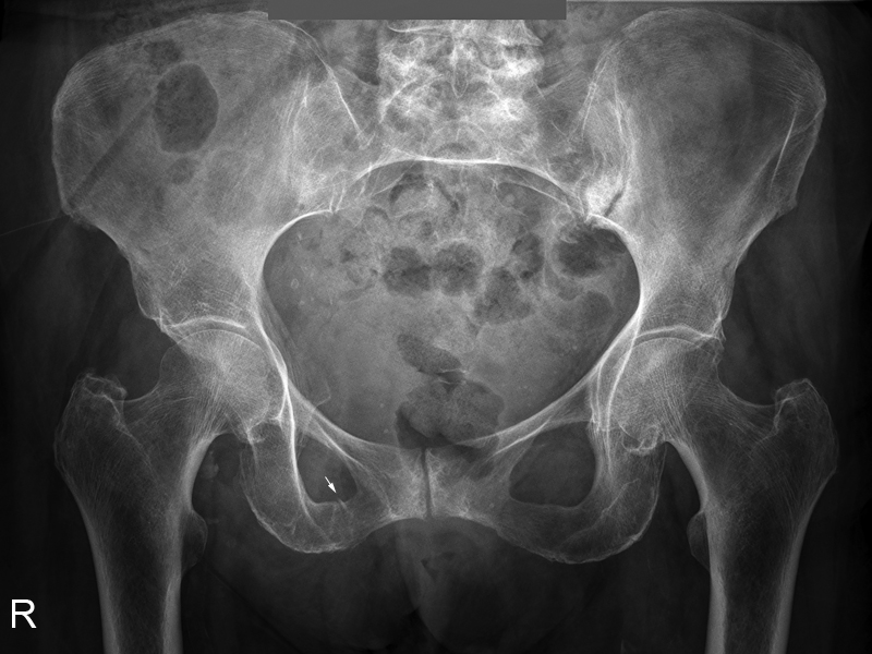
pubic ramiRadiography of Pubic Rami Fractures - wikiRadiographyThe cephalic angle pubic rami projection image provides further support for a fracture of the inferior pubic ramus (arrowed). The slightly sclerotic margin to the fracture is likely to reflect bony overlap rather than evidence of an old fracture.
pubic rami[Untitled]The caudal angle pubic rami projection image does not demonstrate the fracture of the right inferior pubic ramus. The asymmetry of the obturator foramii may be attributed to pelvis rotation, old fracture, or both.


Case 3
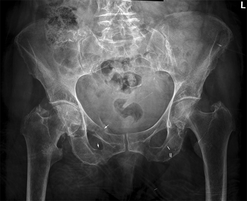
AP pelvisThis 85 year old lady presented to the Emergency Department following a fall onto her left hip. She was examined and was found the be unable to weight bear. She was referred for radiography of her pelvis.

The AP pelvis demonstrates fractures of the right sided pubic rami.

There are a considerable number of artifacts and calcifications
  • metallic sutures
  • metallic vascular clips
  • vascular calcifications
  • blanket artifact
pubic ramiRadiography of Pubic Rami Fractures - wikiRadiographyThe cephalic angled pubic rami image demonstrates the inferior and superior pubic rami fractures
pubic rami[Untitled]The caudal angle pubic rami view image demonstrates the displacement of the superior pubic rami fracture particularly well.


Case 4
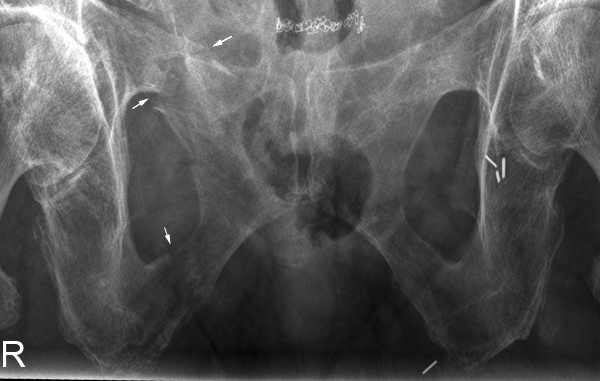
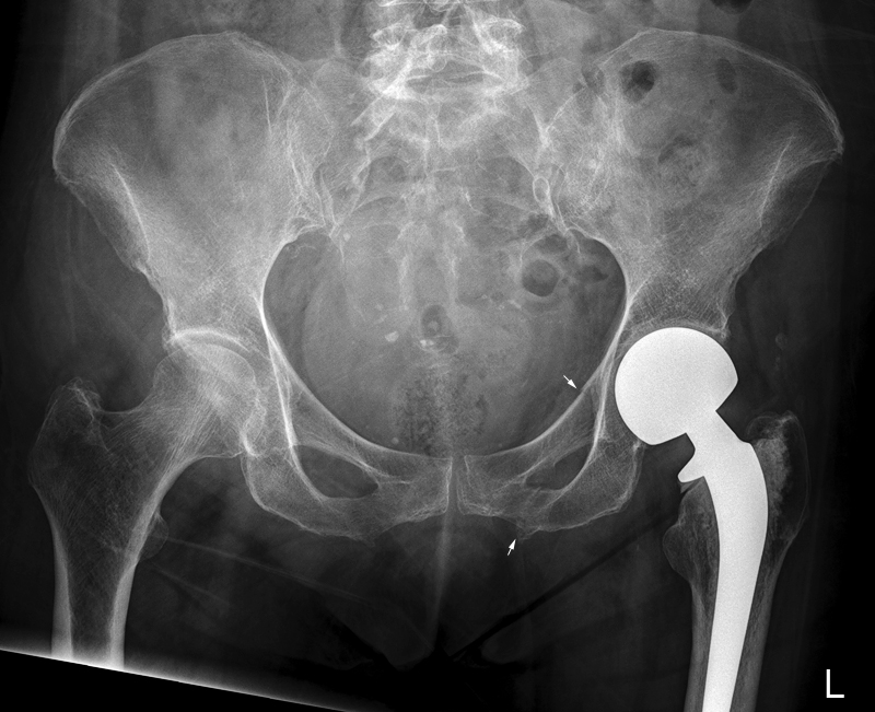
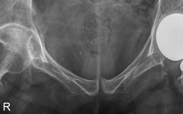
AP pelvisThis 74 year old male presented to the Emergency Department following a fall at the nursing home. He was examined and referred for pelvis radiography with a provisional diagnosis of peri-prosthetic fracture.

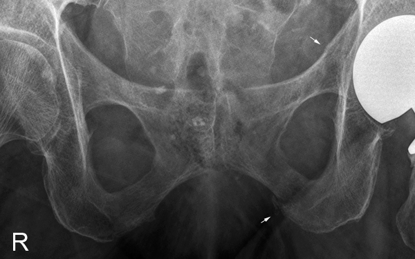
The radiographer noted the presence of a subtle minimally displaced superior pubic ramus fracture (arrowed). There was also a suggestion of an inferior pubic ramus fracture (arrowed). Care must be taken with apparent fractures in this region of the inferior pubic ramus- normal anatomy mimicking a fracture is a common occurrence in this area.
pubic rami[Untitled]The caudal angle pubic ramus projection image did not demonstrate the fracture(s).
pubic ramiRadiography of Pubic Rami Fractures - wikiRadiographyThe cephalic angle pubic ramus projection image demonstrates the subtle superior pubic ramus fracture . The arrowed area of the inferior pubic ramus may represent a fracture, but the appearance is complicated by the overlying soft tissues.


... back to the Wikiradiography home page
... back to the Applied Radiography page