Pelvic Trauma Radiography
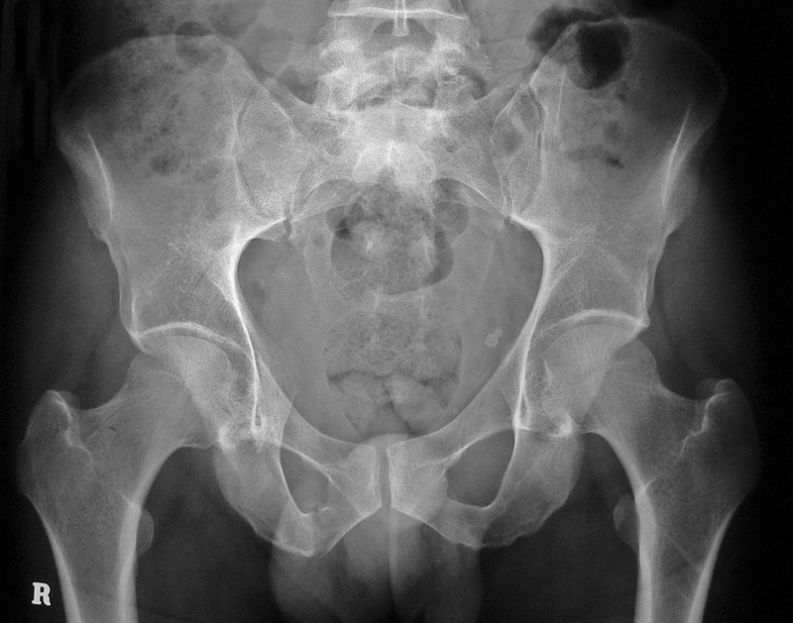
This 44 year old male has presented to the Emergency Department following a motor vehicle accident .
He has severe pelvic pain, particularly on the right side. The patient was referred for a pelvis X-ray examination.
Has the patient suffered any bony injury as a result of his accident? What supplementary views may be useful?
Imaging
Findings
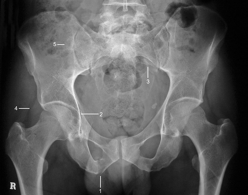
There is a fracture of the right inferior pubic ramus (1). Pubic ramus fractures commonly occur in pairs. It is also noteworthy that there is asymmetry of the obturator foramen. The suspicion of a right superior pubic ramus fracture is very high. On close examination, there is a lucency lying medial to the right acetabulum (2).
This is indeed the expected fracture of the right superior pubic ramus.
In much the same way as you would expect paired fractures to occur in the pubic bones, the pelvis also approximates a ring of bone and satisfaction syndrome should not prevent further examination of the bony pelvis. Fractures of the sacrum are the most commonly missed fractures in pelvic trauma and warrant close examination. The sacral foramen should appear with continuous smooth cortical margins. On close examination, there is a cortical step in one of the left sacral foramen which is almost certainly part of a larger sacral fracture (3)
The soft tissue outlines adjacent to the hip are asymmetrical. This is an unreliable sign of hip injury (4).
The radiographer performed Judet’s views to provide further information as to the nature and extent of the acetabular component of the right superior pubic ramus fracture. Interestingly, these views hid the fracture spectacularly. This is not to decry the value of the Judet’s views, but rather to acknowledge that fractures can occur in almost any plane and that Judet’s views will not always provide new information.
A CT examination of the patient’s pelvis was performed confirming the plain film findings. In addition, a well defined lesion with sclerotic margins was seen adjacent to the right sacro-iliac joint(5).
The fractured sacrum was not as clearly revealed on the CT pelvis images (note circa 1997 CT scanner). The radiographer identified the sacral fracture and performed a dedicated AP sacrum view which, like the Judet’s views, did not help visualize the sacral fracture
Discussion
Dr Lee Rogers has developed the “ring bone paired fractures rule’ into what he has described, with great wit, as the Pretzel-bagel Spectrum. If you <a class="external" href="http://www.google.com" rel="nofollow" target="_blank">google</a> Pretzel-bagel Spectrum all will be revealed.
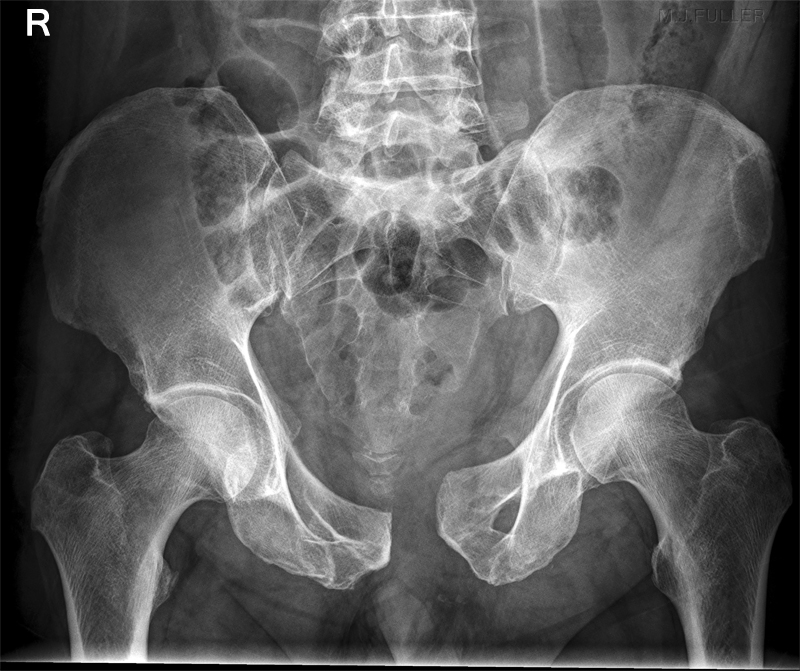
This 69 year old male presented to the Emergency Department following pedestrian vs car trauma. He has right sided pelvic pain and a shortened but not rotated right leg. The patient was referred for a pelvis X-ray examination.
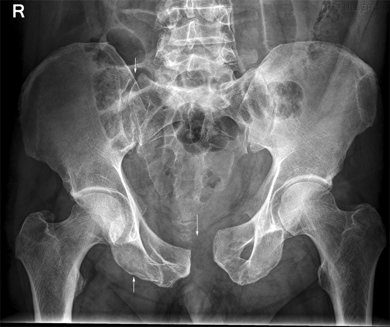
Has the patient suffered any bony injury as a result of his trauma? What supplementary views may be useful? There is diastasis of the symphysis pubis and associated diastasis of the right sacro-iliac joint. There is also a subtle fracture of the right inferior pubic ramus. The sacral arcuate lines (sacral foramina) appear intact.
Potentially useful supplementary views would include pubic ramus views and Judet's views.
No supplementary plain film views were performed- a CT pelvis was performed in lieu of further plain film imaging. In addition to the fractures/subluxations identified on the plain film pelvis image, a comminuted anterior column fracture of the right acetabulum was demonstrated on CT imaging. It is not uncommon for CT imaging of pelvis trauma to reveal new fractures or further information about fractures which have already been identified on plain film imaging.
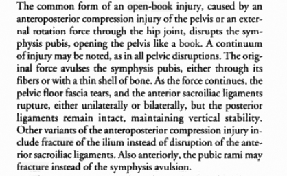
This type of fracture is termed an open book fracture.<a class="external" href="http://books.google.com.au/books?id=OiuuUm8hDfUC&pg=PA173&lpg=PA173&dq=open+book+fracture&source=bl&ots=q-P1B-AZa3&sig=Ry_mgZMFj3Up7N4Q6eYF18yOT70&hl=en&ei=Y8k4Ss-hMsOMkAXC_t2cDQ&sa=X&oi=book_result&ct=result&resnum=14" rel="nofollow" target="_blank">Marvin Tile, David Helfet, James Kellam</a><a class="external" href="http://books.google.com.au/books?id=OiuuUm8hDfUC&pg=PA173&lpg=PA173&dq=open+book+fracture&source=bl&ots=q-P1B-AZa3&sig=Ry_mgZMFj3Up7N4Q6eYF18yOT70&hl=en&ei=Y8k4Ss-hMsOMkAXC_t2cDQ&sa=X&oi=book_result&ct=result&resnum=14" rel="nofollow" target="_blank">Fractures of the pelvis and acetabulum, 2003, p 173</a>
....back to the applied radiography home page here
