Patterns of Consolidation

From wikiRadiography
Revision as of 17:26, 11 November 2020 by Travis (talk | contribs) (Created page with "<div class="WPC-editableContent"><font size="5"><b>Introduction</b></font><br/><blockquote>For those with an interest in plain film image interpretation, patterns of collapse...")
(diff) ← Older revision | Latest revision (diff) | Newer revision → (diff)
Jump to navigation Jump to search
Introduction

For those with an interest in plain film image interpretation, patterns of collapse and consolidation are a very good place to start learning. Plain film chest interpretation is something of a holy grail for radiographers. It might appear to be too difficult to contemplate, but as with most seemingly insurmountable tasks, if you take it a step at a time, you will succeed. This page provides an introduction to the topic.


The Meaning of the Term 'Consolidation'

One of the unfortunate aspects of the term consolidation is that its meaning can be different depending on who is using the term. When a clinician uses the term consolidation, he/she is usually referring to a consolidation associated with acute pneumonia. Thus, the term consolidation and pneumonia have very similar meaning and are almost used interchangeably. Strictly speaking, the term consolidation does not imply any particular aetiology or pathology. Acute pneumonia is the commonest cause but not the only cause of consolidation. (other causes include chronic pneumonia, pulmonary oedema and neoplasm). Thus when a radiologist has reported a chest X-ray examination and notes the presence of consolidation he/she is simply stating that some of the lung airspace has been replaced by a fluid. Sutton, Textbook of Radiology, 2nd ed.,1975,


<embed height="350" src="http://widget.wetpaintserv.us/wiki/wikiradiography/widget/youtubevideo/931dd550c878e17f48c9b80af2d27f80336fa8a8" type="application/x-shockwave-flash" width="425" wmode="transparent"/>


This is a basic video that explains consolidation in simple terms


Notes
  • The term consolidation, when used by a radiologist, does not imply any particular aetiology or pathology. Acute pneumonia is the commonest cause but not the only cause of consolidation. (other causes include chronic pneumonia, pulmonary oedema and neoplasm)
  • Consolidation may be complete or incomplete
  • The distribution of the consolidation can vary widely.
  • A consolidation could be described as “patchy”, “homogenous”, or generalised”.
  • A consolidation may be described as focal or by the lobe or segment of lobe affected

Sutton, Textbook of Radiology, 2nd ed.,1975, p325


Silhouette Sign

Silhouette sign is possibly the most important sign in localising a consolidation and is similarly important in identifying the presence of lung consolidation. The visibility of the borders of thoracic structures such as the heart and diaphragm is dependent on the presence of adjacent air-filled lung. This is not a tricky or sophisticated concept- airfilled lung adjacent to a soft tissue structure like the heart will result in a sharp, clearly defined border between the two structures. This situation may not occur in the presence of any lung disease which causes the airspaces of the lung to fill with fluid- the heart border adjacent to the diseased lung may become obliterated if there is no adjacent air-filled lung to define it.


Anatomy

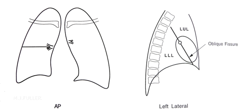
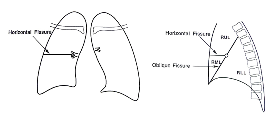
Patterns of collapse and consolidation can definitely not be learned without learning lung anatomy first.

Left Lung

lung anatomy
The left lung has
•1 fissure
•2 lobes


Right Lung

lung anatomy
The right lung has
•2 fissures
•3 lobes


Plain Film Appearances of Lung Consolidation

Radiological appearances common to all lobes are:

1.Abnormal lung opacity
2.Increase in the size and number of lung markings
3.Loss of clarity of the diaphragm on the AP and/or lateral views
4.Loss of clarity of the heart border on the AP and/or lateral views
5.Air bronchogram lines
6.Loss of the normal darkening inferiorly of the thoracic vertebral bodies on the lateral view
7.Opacification of the lung behind the heart shadow or below the diaphragms


1.Abnormal lung opacity------------>
.
2.Increase in the size and number of lung markings
.
3.Loss of clarity of the diaphragm on the AP and/or lateral views
.
4.Loss of clarity of the heart border on the AP and/or lateral views--------->
.
5.Air bronchogram lines
.
6.Loss of the normal darkening of the thoracic vertebral bodies on the lateral view
.
7.Opacification of the lung behind the heart shadow


Notes

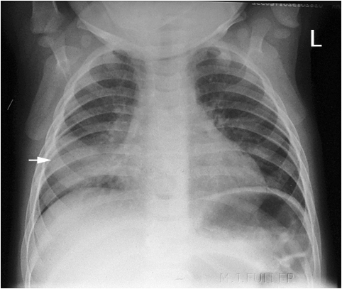
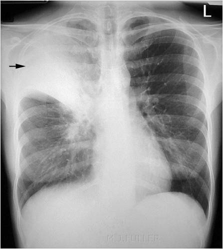
There is abnormal opacity on the right (arrowed). There is also loss of clarity of the right heart border known as silhouette sign.

RML consolidation


1.Abnormal lung opacity -------------->
.
2.Increase in the size and number of lung markings
.
3.Loss of clarity of the diaphragm on the AP and/or lateral views----->
.
4.Loss of clarity of the heart border on the AP and/or lateral views
.
5.Air bronchogram lines
.
6.Loss of the normal darkening of the thoracic vertebral bodies on the lateral view ----------->
.
7.Opacification of the lung behind the heart shadow

Notes

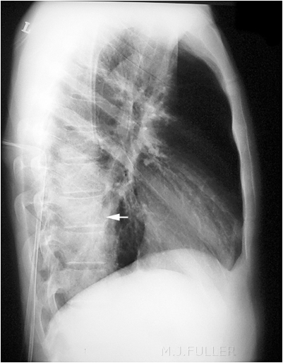
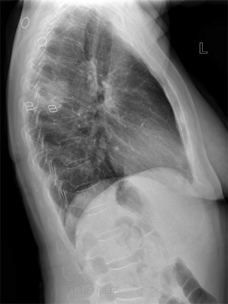
On a lateral chest X-ray image, the thoracic vertebral bodies should appear to darken evenly as you look down the image. This image shows the vertebral bodies become lighter as you move down the image (arrowed). This is caused by consolidation within the left lower lobe. Note also that the right hemidiaphragm is clearly seen and the left hemidiaphragm is only visualised anteriorly (silhouette sign)

Benjamin Felson (<a class="external" href="http://www.amazon.com/Chest-Roentgenology-Benjamin-Felson/dp/0721635911/ref=sr_1_2?ie=UTF8&s=books&qid=1252240078&sr=1-2" rel="nofollow" target="_blank">Chest Roentgenology, W.B. Saunders, 1973, p24</a>) notes that "Equal or greater density of the lower vertebal bodies indicates a pathologic process, even if it is otherwise invisible...".

LLL Consolidation


1.Abnormal lung opacity--------------->
.
2.Increase in the size and number of lung markings
.
3.Loss of clarity of the diaphragm on the AP and/or lateral views
.
4.Loss of clarity of the heart border on the AP and/or lateral views----->
.
5.Air bronchogram lines----------->
.
6.Loss of the normal darkening of the thoracic vertebral bodies on the lateral view
.
7.Opacification of the lung behind the heart shadow

Notes

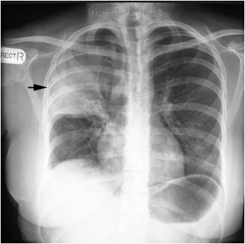
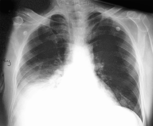
There is a dense opacity within the right upper lobe of the lung (arrowed). There are also air-bronchogram lines and the horizontal fissure has been pulled up by partial collapse of the right upper lobe. There is some loss of definition of the upper right heart border (silhouette sign).

RUL collapse and consolidation


1.Abnormal lung opacity---------->
.
2.Increase in the size and number of lung markings------------>
.
3.Loss of clarity of the diaphragm on the AP and/or lateral views
.
4.Loss of clarity of the heart border on the AP and/or lateral views----->
.
5.Air bronchogram lines----------->
.
6.Loss of the normal darkening of the thoracic vertebral bodies on the lateral view
.
7.Opacification of the lung behind the heart shadow

Notes

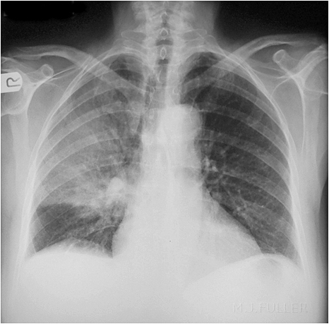
There is abnormal opacity within the right upper lobe. There are air-bronchogram lines. There also appears to be an increase in the number of lung markings, particularly in the peri-hilar region of the right.

RUL consolidation


1.Abnormal lung opacity---------->
.
2.Increase in the size and number of lung markings
.
3.Loss of clarity of the diaphragm on the AP and/or lateral views
.
4.Loss of clarity of the heart border on the AP and/or lateral views
.
5.Air bronchogram lines------------>
.
6.Loss of the normal darkening of the thoracic vertebral bodies on the lateral view
.
7.Opacification of the lung behind the heart shadow---------->

Notes

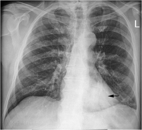
There is abnormal opacity behind the left heart shadow (arrowed). There are also air-bronchogram lines and loss of the left paraspinal stripe


LLL Consolidation


Right Upper Lobe (RUL) Consolidation
RUL consolidation
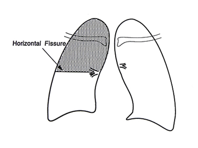
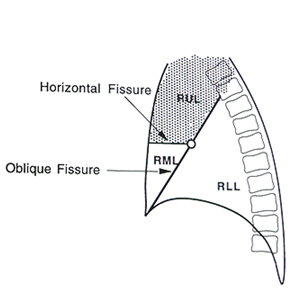
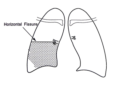
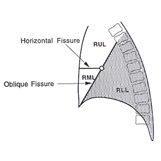
RUL consolidation will be seen as an increased opacity within the shaded area. Opacity may be sharply bordered by the horizontal fissure
Some loss of outline of the upper right heart border may be apparent
RUL consolidation
  • Dense opacity seen above the horizontal fissure.
  • Air-bronchogram line
RUL consolidation
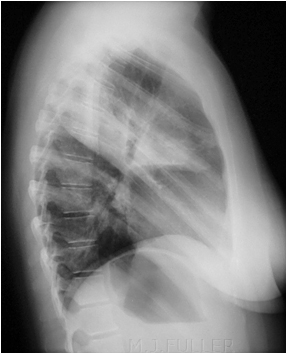
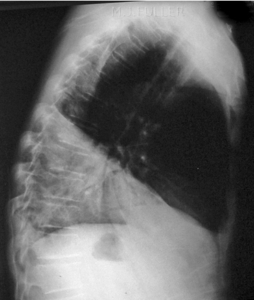
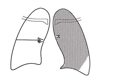
In the lateral view, there will be increased density in the RUL which may be sharply bordered by the horizontal and/or oblique fissure(s).
RUL consolidation
  • Dense opacity in the RUL sharply bordered by the horizontal and oblique fissures


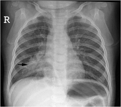
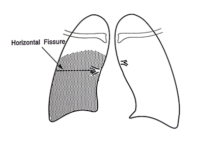
Right Middle Lobe (RML) Consolidation
RML consolidation
•Seen as an area of increased opacity in the shaded area
•Loss of the definition of the right heart border is often seen

RML consolidation
  • RML opacification
  • Loss of adjacent right heart border
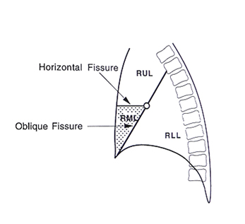
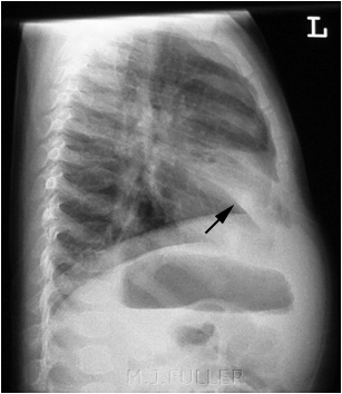
RML consolidation
•RML consolidation is characteristically seen as a wedge opacity in the lateral view
•May be sharply bordered by the horizontal and oblique fissures
•(collapse of the lingula segment of the LUL has a similar appearance)
RML consolidation
  • Wedge shaped opacity characteristic of RML consolidation(black arrow)
  • lingula segment consolidation can have a similar appearance on the lateral view
  • some RML collapse also present


Right Lower Lobe (RLL) Consolidation
RLL consolidation
•Appears as an area of increased opacity within the RLL
•Some loss of the hemi-diaphragm is commonly seen
RLL consolidation
•Loss of right hemi-diaphragm
•Dense opacity in RLL
•Some loss of right heart border
RLL consolidation
•Increased opacity within the RLL
•Commonly seen with loss of the right hemi-diaphragm
RLL consolidation
•Triangular opacity
•Loss of right hemi-diaphragm


Left Upper Lobe (LUL) Consolidation


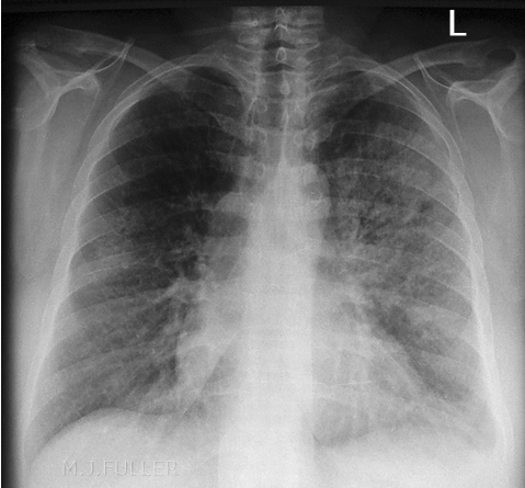
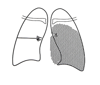
LUL CONSOLIDATION
•Appears as an area of increased opacity within the LUL
•Characteristically not a dense opacity on the PA view
•Often loss of the upper mediastinal contour
LUL CONSOLIDATION
•Opacity left hemi-thorax
•Air-bronchogram lines
•Some loss of left heart border
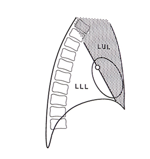
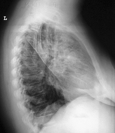
LUL CONSOLIDATION
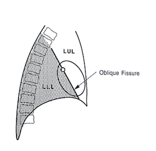
•Can be sharply bordered by the oblique fissure
•Does not involve the diaphragm
LUL CONSOLIDATION
•Opacity seen anterior to the oblique fissure

Left Lower Lobe (LLL) Consolidation


LLL Consolidation
•Appears as an area of increased opacity within the LLL
•Some loss of the hemi-diaphragm is commonly seen
•May be increased density behind left heart shadow
LLL Consolidation
•Appears as an area of increased opacity within the LLL
•Some loss of the hemi-diaphragm medially is seen
•increased density behind left heart shadow
LLL Consolidation
•Increased opacity within the LLL
•Commonly seen with loss of the Left hemi-diaphragm
•May be sharply delineated by oblique fissure
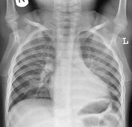
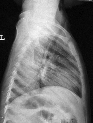
LLL Consolidation
•Increased opacity within the LLL
•Loss of the normal darkening of the t spine some loss of the left hemi-diaphragm posteriorly


Round Pneumonia
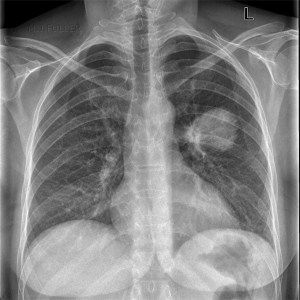
A special case of consolidation is known as round pneumonia. This is where there has been a focus of infection in the lung with local spread.
round pneumoniaround pneumonia
This patient presented with pyrexia of unknown origin (PUO). There is a dense opacity in the mid/upper region of the left lung. In a patient who presents with pyrexia, round pneumonia must be considered.

Neoplasm cannot be excluded but is unlikely. There is a suggestion of an airbronchogram within the lesion. This excludes a solid tumour and also excludes a cystic lesion.
The lesion is shown to be posterior, possibly in the superior segment of the lower lobe.


...back to the Applied Radiography home page