Orthopaedic Screws,Plates and Prosthesis
Jump to navigation
Jump to search
under construction
Introduction
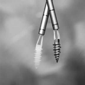
Tendon ScrewsA knowledge of orthopedic devices and how they are used can be useful to the radiographer. Old imaging is not always available at orthopedic clinic. If the patient tells you they have a "hook plate", it is useful to know what they are talking about in order to approach the radiography with confidence.
It is widely considered good practice to include all of an orthopedic device during follow up radiography. A common problem is to know how far the device extends along the diaphysis of a long bone. This page is aimed at providing examples of common orthopaedic devices.

Source: Smith and Nephew in <a class="external" href="http://www.oag-bvg.gc.ca/internet/English/att_20040302pe03_e_13197.html" rel="nofollow" target="_blank">http://www.oag-bvg.gc.ca/internet/English/att_20040302pe03_e_13197.html</a>