Orthopaedic Clinic Wrist Radiography
There are significant differences in radiography provided in an acute setting to a non-acute setting. In large hospitals, most of the acute orthopaedic radiography will occur in the X-ray rooms which service the Emergency Department. Patients who have orthopaedic injuries requiring follow-up will be seen at orthopaedic clinic and they are usually referred for follow-up radiography of their injury. This page considers wrist radiography techniques that are more commonly employed in follow-up orthopaedic examinations.
Why Follow-up Treated Orthopaedic Injuries?
The simple answer to this question is that there can be a variety of unexpected outcomes including non-union and changes in fracture alignment. A patient with a healed fracture can have immobilisation/support devices removed.
Case 1
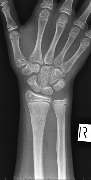
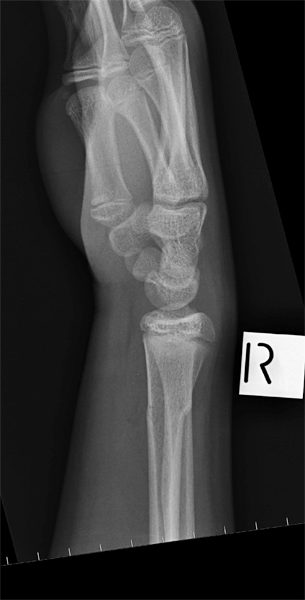
This 14 year old boy presented to the Emergency Department after falling onto an outstretched hand. He had a painful and swollen wrist and was referred for right wrist radiography.
There is a fracture of the distal radius which appears undisplaced in this view. The fractured distal radius is demonstrated on the lateral view with minor dorsal angulation.
The patient received a fibreglass plaster and arrangements were made for the patient to return to orthopaedic clinic in 2 weeks to assess the healing of the fracture.
CommentIsolated fractures of the distal radius tend to displace late and get stuck <a class="external" href="http://www.orthospot.com.au/papers.orthospot.com.au/fracupl_files/frame.htm" rel="nofollow" target="_blank">(http://www.orthospot.com.au/papers.orthospot.com.au/fracupl_files/frame.htm)</a>. This plaster shows a lack of mould points which may have precipitated the angular displacement of the distal radius fracture.
... back to the Wikiradiography home page
... back to the Applied Radiography page


