Orthopaedic Clinic Radiography

From wikiRadiography
Revision as of 17:22, 11 November 2020 by Travis (talk | contribs) (Created page with "<div class="WPC-editableContent"><b><font size="5">Introduction</font></b><br/><blockquote>Radiographers who are employed in larger hospials will see patients who are referred...")
(diff) ← Older revision | Latest revision (diff) | Newer revision → (diff)
Jump to navigation Jump to search
Introduction

Radiographers who are employed in larger hospials will see patients who are referred from outpatient consulting clinics. Usually, a large number of these patients will be referred from orthopaedic clinic for radiography of fractures diagnosed in the Emergency Department (and elsewhere). This tends to be repetitive work and the objectives of re-imaging these fractures and dislocations may not always be clear. This page considers all aspects of orthopaedic clinic radiography.


Case 1
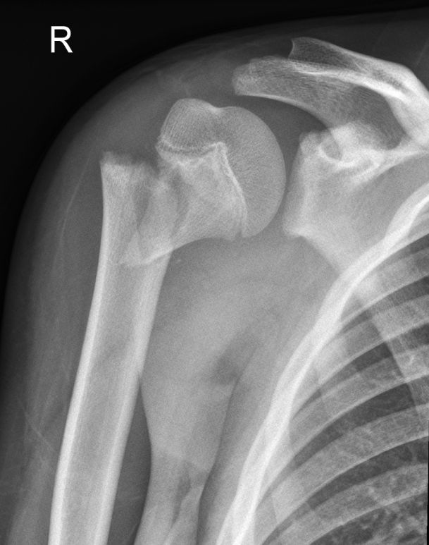
SHII prox humerusThis 10 year old boy presented to the Emergency Department following a fall from his pushbike. He was examined and found to have a swollen and painful right shoulder. He was referred for right shoulder radiography.

This is an AP shoulder image with the patient's arm in internal rotation. The radiography is compromised to suit the patient's condition. There is a proximal humerus fracture (? Salter-Harris II)
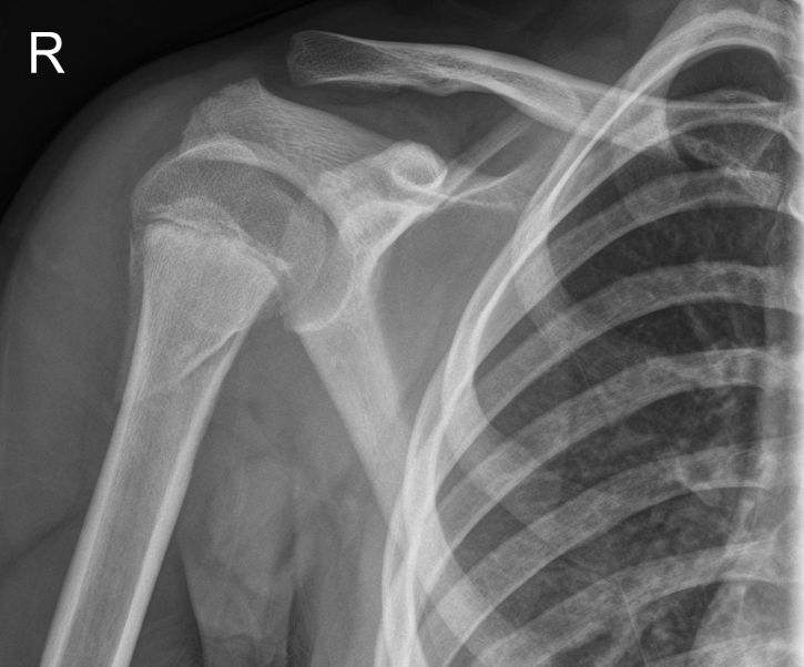
AP ahoulderThe patient was treated with a collar-and-cuff and referred to orthopaedic clinic for follow-up assessment of the injury. With the patient now in a less distressed state, the radiographer was able to achieve a true AP shoulder with external rotation of the humerus. The distal fragment is now demonstrated with significant displacement. Note that the humeral position is not comparable with the original position as demonstrated in the Emergency Department imaging. It is worth considering the need to provide radiographic demonstration of these fractures in true AP and lateral positions in the Emergency Department so that follow up imaging will be comparable. It is conceivable that if the two positions of the humerus are at 90 degrees (orthogonal), there may be no change in the actual position/displacement of the fracture i.e. apparent displacement is positional rather than actual.