Odontoid-lateral mass Asymmetry
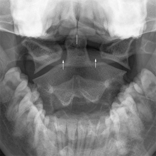
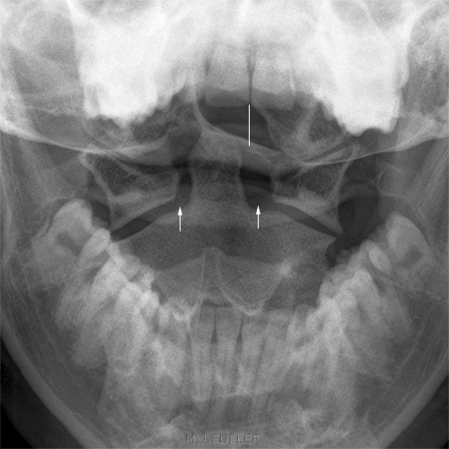
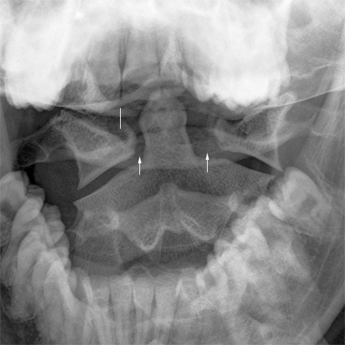
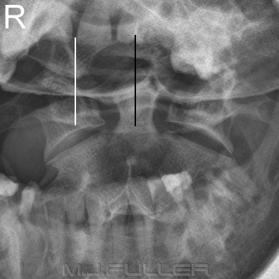
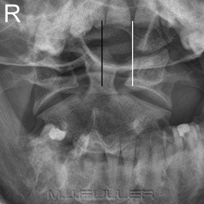
Asymmetry of the lateral masses of C1 with respect to the odontoid process is commonly seen on the AP C1/C2 image. The difficulty with this finding in trauma patients is to know what to do next. The finding is very commonly found to be a normal variant or idiopathic. It does not however seem appropriate to ignore the finding in trauma patients.
Research
Odontoid-lateral mass asymmetry in minimally symptomatic patients: a normal variant. <a class="external" href="http://www.springerlink.com/content/101157/?p=36a71e9098224d31ac38ea9529bc8e93&pi=0" rel="nofollow" target="_blank">European Journal of Orthopaedic Surgery & Traumatology. </a><a class="external" href="http://www.springerlink.com/content/e3u1acd3gj5u/?p=36a71e9098224d31ac38ea9529bc8e93&pi=0" rel="nofollow" target="_blank">Volume 13, Number 2 / June, 2003</a>
Case 1 Discussion
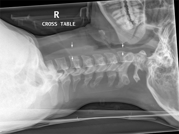
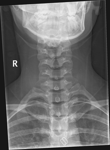
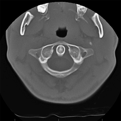
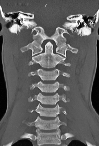
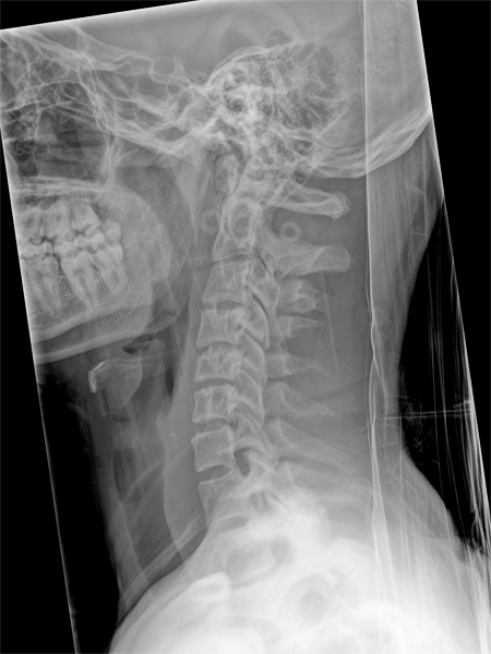
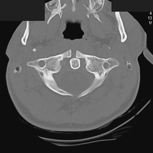
You could speculate as to the cause of the apparent odontoid-lateral mass asymmetry on the initial plain films. I would suggest that it was not purely projectional- three slightly different AP projections demonstrated the same asymmetry. Was it caused by the soft tissue injury that the patient received during the trauma? Was it associated with the wearing of the cervical hard collar? Was it a combination of a number of factors? Importantly, the asymmetry was not apparent on CT scan imaging 24 hours after the initial trauma. This case does not support the notion of asymmetry as a normal variant- the asymmetry was not evident 24 hours after the injury.
It is noteworthy that the head rotation employed was minimal and may not have been sufficient to produce appreciable difference in the lateral mass-dens distances.
Case 2
Case 2 Discussion
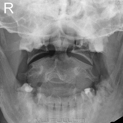
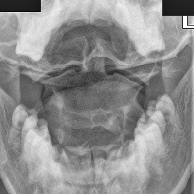
The LPO position in particular shows evidence of symmetry of the C1 lateral masses with respect to the dens and with respect to the body of C2. This additional information needs to be considered in the context of:
- clinical signs (e.g. paresthesia, head control, pain)
- mechanism of injury (e.g. high speed MVA)
- evidence of injury demonstrated on the other cervical spine projections
- soft tissue signs of injury.
Case 3
Discussion
These cases demonstrate a variety of possible findings associated with odontoid-lateral mass asymmetry. It is useful for the radiographer to be aware of the significance of this appearance and the possible imaging options and diagnostic outcomes
... back to the Applied Radiography home page

