Oblique Lumbar Spine Technique
AnatomyOblique projections of the lumbar spine are now infrequently performed in some centres. The diagnostic yields from this view are considered to be limited and the radiation dose to the patent is significant. CT scanning is a more accurate diagnostic tool but is also a high radiation dose procedure.
Libson et al (Radiology 1984; 151: 89-90) argued in 1984 that the oblique view was still important and that defects in the pars interarticularis (spondylolysis) could be missed if oblique views were not performed. Libson's research found that of the patients that demonstrated spondylolysis, 20% were revealed on the oblique views only.
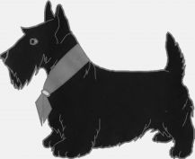
This is the RPO or LAO position to show the lower lumbar vertebra (without red outline) The red outlined anatomy is commonly known as "Scotty Dog".


 source: unknown | [[1]] |
The spinal facet joints vary in their orientation both with age and between anatomical regions. Some radiographers with perform lateral lumbar spine with the patients upper lumbar spine at a less acute angle than the lower lumbar spine. The success of this position may be accounted for by consideration of the changes in the angles of the facet joints as you move proximally up the lumbar vertebrae.

Pars Defects- Traumatic or Congenital?
John Harris notes (The Radiology of Emergency Medicine, 3rd Ed, 1993, p 273, Williams and Wilkins) "The etiology of spondylolysis is overwhelmingly congenital, although sporadic reports of traumatic lumbar spondylolysis appear in the literature". CT imaging will usually identify the defects as characteristically acute or otherwise.
Positioning Technique
Oblique lumbar spine radiography can be performed PA or AP. The advantage of the PA approach is ease of positioning. It is easier to centre to the spine when surface anatomy landmarks provide a clear guide. The AP approach is arguably more difficult to achieve but has the advantage of being easier for the patient and the anatomy is closer to the IR resulting in improved image quality. Some centres will not require the upper lumbar vertebra to be included- the reason for this is that the view is usually performed for pars interarticularis defects which occur almost exclusively at the lower lumbar levels (in acute injury patients).
Tip- when using the PA approach, position the patient with their shoulders at a shallower angle than their hips...based on experience rather than science
- Which way do I oblique the patient?
- RPO and LAO show right pars interarticularis, LPO and RAO show left pars interarticularis
- both obliques are usually performed
- How much do I oblique?
- 30 - 45 degrees (varies from 30 to 50 degrees between patients, and may not be the same on both sides
- What is the anatomy demonstrated?
- the pars interarticularis is usually the anatomy of interest. Facet joint degeneration can also be demonstrated
- AP or PA?
- your choice- consider image quality issues, patient's abilities
- How do you label the images?
- label the side where the facet joint is in profile (see below)- for supine/AP technique it is the side down, for prone/PA technique it is the side up
- Where do I centre?
- if you are aiming to demonstrate the entire lumbar spine, centre at the level of the lower costal margin centred to the spine
- for lower lumbar vertebra, centre at the level of the ASIS
- don't make the mistake of centring over the spinous process when using the PA approach (look at the axial anatomy of a lumbar vertebra)
- for supine technique, the centre point is 2 inches (5cm) medial to the ASIS
- What FFD/SID should I use?
- 40 inches or 115cm
- How much do I angle the beam?
- depends whether you are imaging the entire lumbar vertebra or coning to L5S1- for imaging the entire lumbar vertebra, don't angle the beam. For imaging the lower lumbar vertebra (L4/5, L5/S1) angle to match the lumbar lordosis
- also depends on the patient's lumbar lordosis
- What exposure technique should be employed?
- You may need a considerable increase in exposure compared to the AP/PA lumbar spine
- breathing technique could be considered in selected patients but is likely to be of limited benefit
Is Tube Angulation Necessary?
This is a supine lateral cross-table lumbar spine image. The white lines represent the diverging X-ray beam (albeit exaggerated) as might occur with an AP view of the lumbar spine in this patient. It would be expected that the L3/4 intervertebral disc space would be displayed clearly. As you move further away from this central disc space, the discs would appear increasingly less of a true AP.
A similar effect is seen with AP oblique projections of the lumbar spine
Angulation of the beam (caudal or cephalic) would be of little benefit (AP or oblique) given that it would favour some levels and distort others.
Of course, imaging of the intervertebral discs is not important with oblique views of the lumbar spine. However, if the lumbar intervertebral discs are not imaged in profile, imaging of the posterior elements of the spine will also be compromised.
<a class="external" href="http://www.medscape.com/" rel="nofollow" target="_blank" title="http://www.medscape.com">http://www.medscape.com/content/1999/00/40/85/408509/art-mos4931.solo.fig4.jpg
</a>These images demonstrate the tendency to produce an undistorted view of the central lumbar vertebrae in the oblique projection. (white arrow only).
There are two strategies that might help avoid this problem.1. Use a PA technique may help(match lumbar lordosis to divergent rays)
2. Image the lower 3 lumbar discs only and angle the X-ray beam to suit (cephalic angle for AP technique and caudal angle for PA technique)This is a coned AP oblique projection of the lower lumbar vertebra with cephalic angulation. The displayed vertebrae are demonstrated with less distortion than using the 'straight tube' technique above. The Scotty dog profiles are demonstrated clearly because they are imaged without distortion.


Erect Oblique Lumbar spine Technique
Centre Point for PA Technique
Erect AP Oblique Lumbar Spine Erect PA Oblique Lumbar Spine
Note that the centre point is not the spinous process! (see below)


Incorrect Correct This is a trap that you might fall into the first time you attempt an oblique lumbar spine in the prone/PA oblique position. The beam is centred to what appears to be the spine based on surface anatomy This is the correct positioning. Centre onto the raised side by at least an inch (d = 3.5cm). The exact centring point will depend on the patient's body habitus and degree of obliquity


Images
Should you Include all of the Lumbar Vertebrae on Oblique Views?
This is the RPO or LAO position to show the lower lumbar vertebra.
Film/screen technique with round coneThis is the LPO or RAO position to show the lower lumbar vertebra
Film/screen technique with round cone
PathologyLibson et al (Radiology 1984; 151: 89-90) reported the following
Libson et al (Radiology 1984; 151: 89-90)To take the authors argument even further, the diagnostic yield from including L3 will be very small and arguably not worth the cost in terms of patient radiation dose.


When viewing an oblique lumbar spine image, you are most interested in Scotty's ear, leg and neck. Scotty's leg and ear are the superior and inferior articulating processes respectively. In patients with facet joint degeneration, changes will be seen in the scotty's ear and leg. More importantly, scotty's neck is of great interest. If scotty appears to be wearing a white collar, the patient may have a defect in the pars interarticularis manifest as a sclerotic band. If scotty's neck is missing, the patient may have a traumatic injury to the pars interarticularis, a congenital defect or both.
It is important to note that a patient who has a defect of the pars interarticularis (scotty's neck) may not have an acute injury. The defect may be congenital.
<a class="external" href="http://www.gentili.net/signs/20.htm" rel="nofollow" target="_blank">http://www.gentili.net/signs/20.htm</a>
<a class="external" href="http://www.gentili.net/signs/20.htm" rel="nofollow" target="_blank">http://www.gentili.net/signs/20.htm</a>normal Scotty 'No neck' Scotty at L5 (spondylolysis and spondylolisthesis)


Case 1
This 20 year old female patient presented to the Emergency Department with low back pain. The patient had a history of involvement in a motor vehicle accident (MVA) 24 hours prior.
This is an AP lumbar spine image taken with a long exposure time using a film/screen technique. This is a lateral lumbar spine using a long exposure time and long ffd on film/screen . In addition, an aluminium wedge filter has been placed to reduce the exposure to all but L5 and lower. There is a defect of the L5 pars interarticularis- probably bilateral (white arrow). The coned lateral L5S1 image supports the diagnosis of a L5 pars interarticularis defect.
Elastic artifact noted


Comment
The referring doctor reviewed the images and referred the patient for CT lumbar spine imaging in lieu of obliques of the lower lumbar spine. The pars defects were shown to be well corticated suggesting that they were old and probably congenital.
Case 2
This 24 year old female patient presented to the Emergency Department following acute onset of low back pain. The patient had recently been on a long bus trip. The patient has a history of involvement in a motor vehicle accident (MVA) 5 years ago.
The lateral lumbar spine demonstrates pars interarticularis defects at L3 (film/screen technique circa 1997) The coned RPO oblique view demonstrates a pars interarticularis defect on the right Similarly, the left oblique demonstrates a pars interarticularis defect.


Comment
The pars defects in this case were considered to be non-acute.
...back to the Applied Radiography home page