Nutcracker Fracture of the Cuboid

From wikiRadiography
Revision as of 17:21, 11 November 2020 by Travis (talk | contribs) (Created page with "<div class="WPC-editableContent"><div align="left"><font size="5"><b>Introduction</b></font><br/><blockquote>A <b><i>nutcracker fracture</i></b> of the cuboid is a very uncomm...")
(diff) ← Older revision | Latest revision (diff) | Newer revision → (diff)
Jump to navigation Jump to search
Introduction

A nutcracker fracture of the cuboid is a very uncommon fracture. This page considers all aspects of the radiography of the nutcracker fracture


Acknowledgement

I have drawn extensively from the work of Nicholas Beckmann, MD and Manickam Kumaravel, MD (The Department of Diagnostic and Interventional Imaging, The University of Texas Medical School). Their presentation <a class="external" href="http://www.uth.tmc.edu/radiology/presentations/2008/nutcracker_beckman_2008.pdf" rel="nofollow" target="_blank">Nuts and Bolts of the Nutcracker</a> is recommended for further reading.


Anatomy
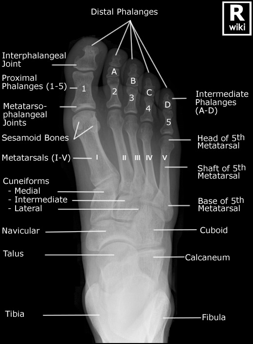
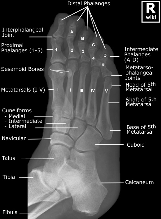
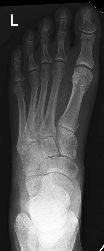
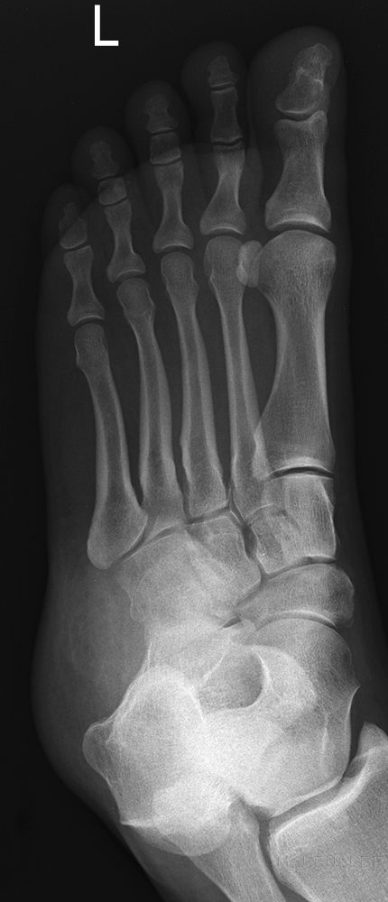
Radiographic Anatomy - Foot DPRadiographic Anatomy - Foot Oblique
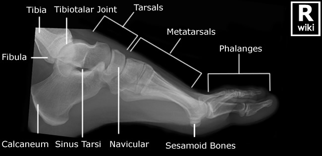
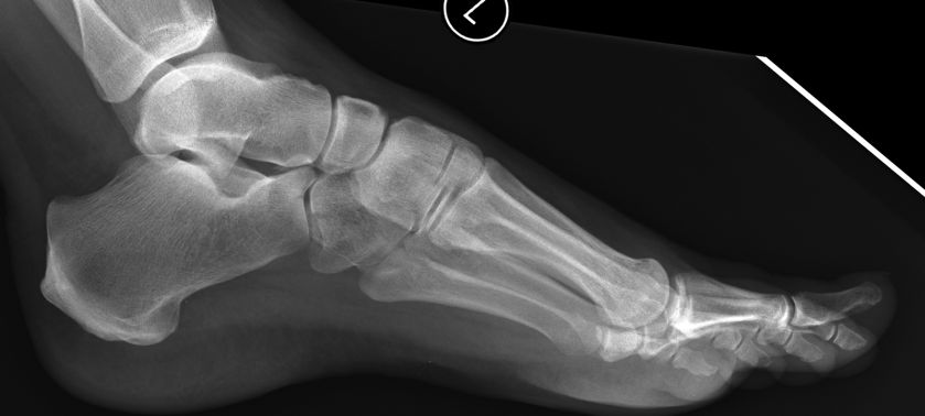
Radiographic Anatomy - Foot Lateral


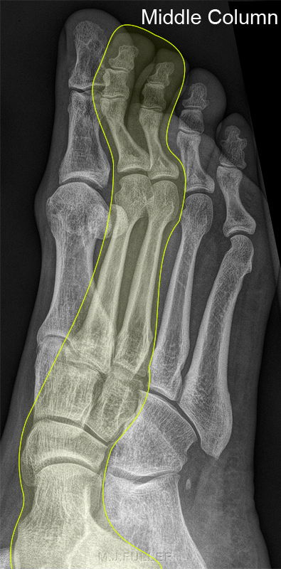
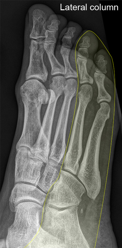
The Columns of the Foot

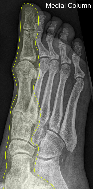
Foot- medial columnMedial Column of the foot

•1st metatarsal
•Medial cunieform
•Navicular
•Talus

Foot- middle columnLateral Column of the foot

•2nd & 3rd metatarsals
•Middle & lateral cunieforms
•Navicular
•Talus

lateral columnLateral Column of the foot

•4th & 5th metatarsals
•Cuboid
•Calcaneus


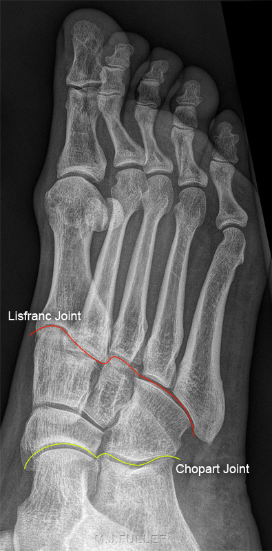
Midfoot Articlations

midfoot articulationsMidfoot articulations with forefoot and hind foot

•Lisfranc joint (forefoot articulation, red line)
•Chopart joint (hind foot articulation, yellow line)


What is a nutcracker Fracture?
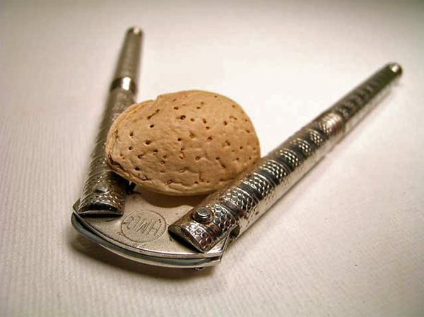
nutcracker
Nicholas Beckmann, MD and Manickam Kumaravel, MD <a class="external" href="http://www.uth.tmc.edu/radiology/presentations/2008/nutcracker_beckman_2008.pdf" rel="nofollow" target="_blank">Nuts and Bolts of the Nutcracker</a> . http://www.uth.tmc.edu/radiology/presentations/2008/nutcracker_beckman_2008.pdf
  • the nutcracker fracture derives its name from the action of a nutcracker which can open a nut by cracking the shell by compressing it between the two handles (see nutcracker left)

•Most cuboid fractures are caused by compression of the cuboid between the calcaneus and the lateral metatarsals during force abduction
•The mechanism similar to cracking a nut, hence called “nutcracker fractures”


Nutcracker Fractures

Case 1

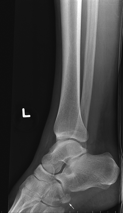
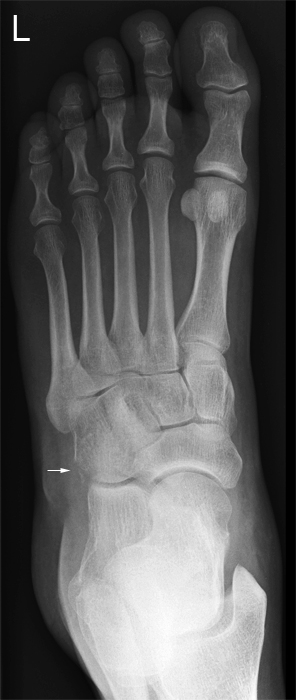
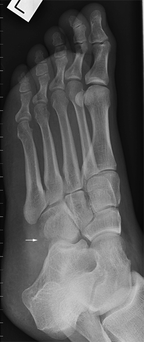
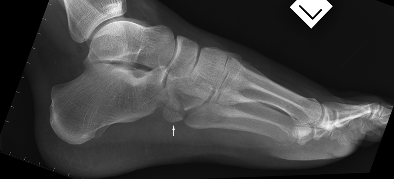
This 50 year old male presented to the Emergency Department following a fall from a ladder. On landing his foot became stuck. He was referred for ankle radiography initially then re-referred for foot radiography.

LATERAL ANKLEThe lateral projection of the ankle demonstrates a fracture of the cuboid (arrowed).


AP footOblique Foot
The DP projection of the foot demonstrates a fracture of the cuboid (arrowed).The fracture of the cuboid appears to be more extensive on the oblique view of the foot (arrowed).
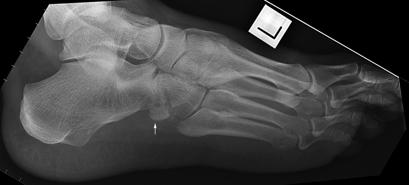
Lateral footThe cuboid fracture is well demonstrated on the lateral foot image and appears to have a large plantar component. The lateral foot position is not true lateral which has tended to demonstrate the lateral column of the foot to advantage. The fracture appears to be a nutcracker fracture of the cuboid.
oblique footFurther obliquity from the lateral position has possibly demonstrated the donor site from the cuboid of the arrowed fragment.


Case 2

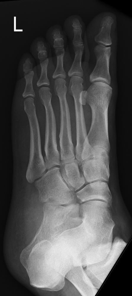
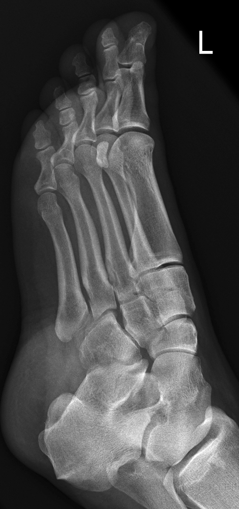
nutcracker fractureThis 42 year old female presented to the Emergency Department after tripping and rolling her left ankle. She was examined and referred for foot radiography.

There is no clearly demonstrated displaced fracture
nutcracker fractureThere is no clearly demonstrated displaced fracture
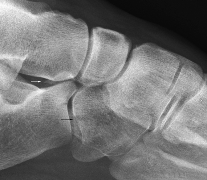
nutcracker fractureThere is a cuboid cortical defect and lucency demonstrated.
nutcracker fractureMagnified image shows cuboid fracture (black arrow) and possible calcaneal fracture.
Nutcracker Fracture of the Cuboid - wikiRadiographyThere is no clearly demonstrated displaced fracture
nutcracker fractureCuboid fracture demonstrated